EN
EN
SD Rat Costal Chondrocytes
Introduction
Cartilage tissue, composed of chondrocytes, matrix, and fibers, possesses distinct regenerative capacity. Within this tissue, proliferating immature cells, resembling fibroblasts, gradually differentiate into chondroblasts and secrete the cartilage matrix. As the cells become embedded in cartilage lacunae, they mature into resting chondrocytes. Immature chondrocytes are distributed individually in the superficial layer of cartilage, characterized by a small, oval shape, with their long axes aligned parallel to the cartilage surface. Toward the deeper layers, the cells increase in size and become rounded, with lightly stained circular or oval nuclei. Their cytoplasm shows weak basophilia, and variable numbers of lipid droplets are frequently observed.
OriCell™ SD Rat Costal Chondrocytes are isolated from rib cartilage tissue via a combined trypsin-collagenase digestion method to ensure high purity. The in vitro culture of these chondrocytes provides a valuable model for investigating their physiological functions, drug responses, and pathophysiological changes under various pathological conditions.
When citing our products in academic publications, please use the following format: "OriCell™ [Product Name] + [Catalog Number], from Cyagen Biosciences."
Product Information
| Product Name | OriCell™ SD Rat Costal Chondrocytes |
| Catalog Number | RASRB-02071 |
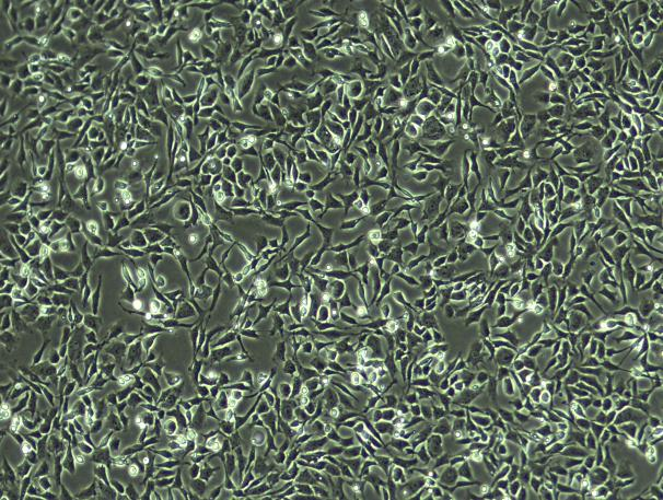
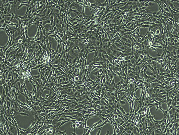
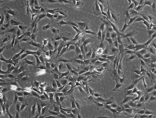
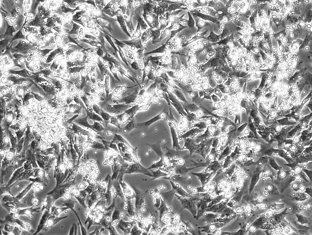
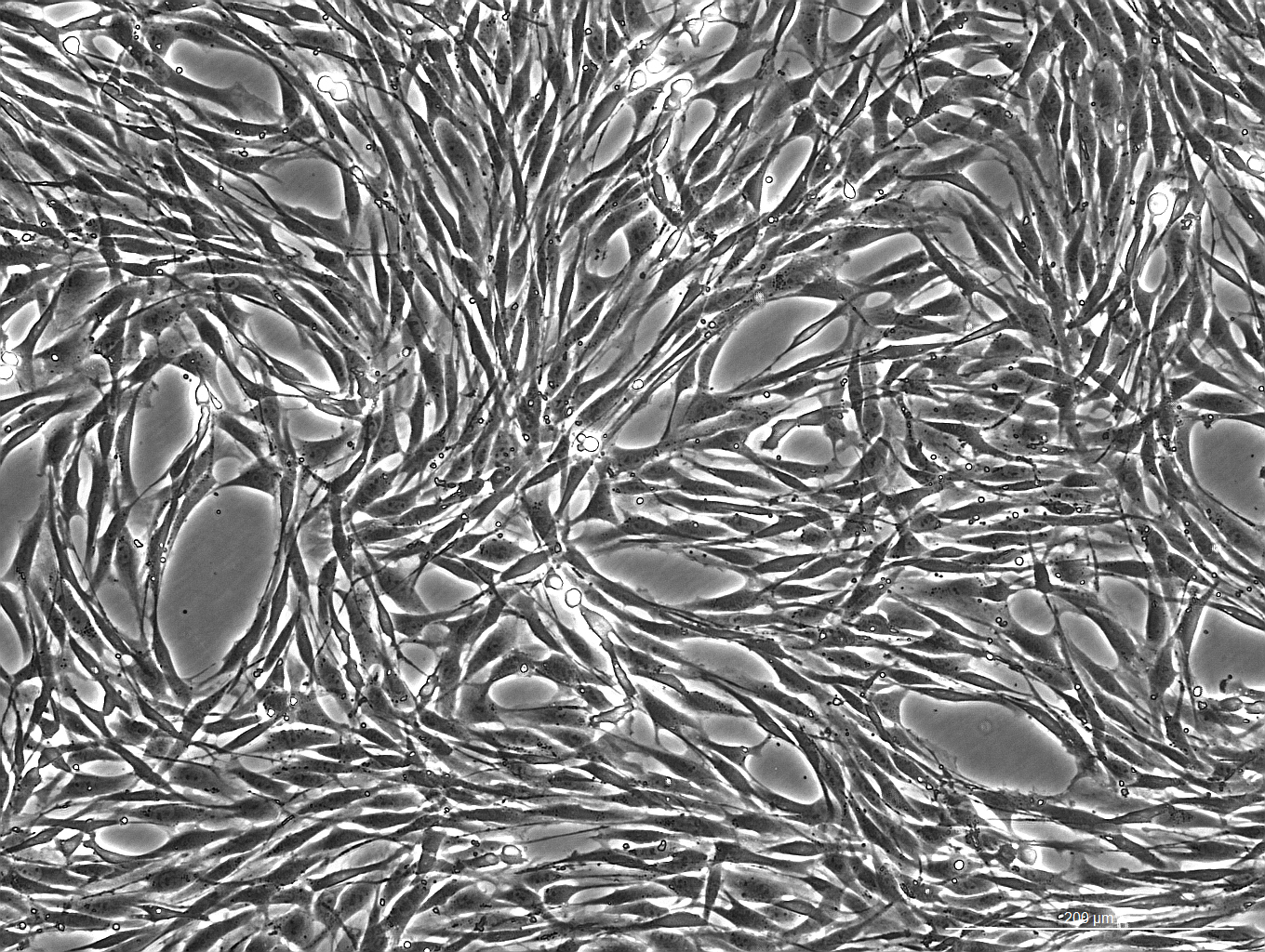
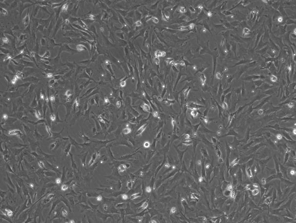
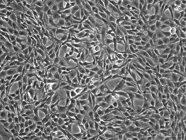
| Cell Characteristics | Adherent growth; Spindle-shaped or triangular-shaped |
| Passage Capability | Limited; Prioritize low-passage cells |
| Identification Markers | Collagen Ⅱ |
| Number of Cells | 1×10⁶ cells/vial |
| Passage Number | P1 |
| Storage at | Liquid Nitrogen (-196℃) |
QC
- Pass the detection of bacteria, fungi, mycoplasma, and endotoxins.
- Verified by cell recovery viability testing, with a post-thaw survival rate >80%.
- Cell identification: Positive for Collagen Ⅱ expression (>80%) as confirmed by immunofluorescence (IF) staining.
Please refer to "COA" for details.
General Handing Principles
- Maintain strict aseptic technique. Ensure complete sterility throughout all procedures, particularly within the laminar flow hood and incubator.
- Follow standardized protocols. Adhere strictly to the product manual. Implement rigorous control over experimental variables and include appropriate parallel controls.
- Use high-quality consumables and reagents. This product requires culture vessels suitable for adherent cell growth, and the reuse of these vessels is not recommended. The reagents used must be validated for reliability, cell compatibility, and batch-to-batch consistency.
- Prioritize low-passage cells. Since OriCell™ SD Rat Costal Chondrocytes have limited ability to proliferate in vitro, we recommend using low-passage cells for research applications.
- Optimize seeding density and subculture. The recommended seeding density for OriCell™ SD Rat Costal Chondrocytes is (2.5–4) × 10⁴ viable cells/cm². Since cell growth is highly dependent on donor characteristics and culture conditions, we recommend adjusting the split ratios based on the actual performance of each specific lot and passage.
Warm Notice: The cryopreservation medium of this product contains DMSO, which may pose potential risks. Please handle it with care.