
How to Identify Mesenchymal Stem Cells (MSCs)
Mesenchymal stem cells (MSCs) are a type of multipotent stem cell derived from the mesoderm. They were first discovered in bone marrow and have attracted sustained attention from researchers ever since. MSCs can be obtained from a wide range of sources, including bone marrow, adipose tissue, dental pulp, placenta, umbilical cord, and liver, among others. Even after multiple passages in culture, they maintain strong proliferative and differentiation potential. In addition, MSCs exhibit low immunogenicity, which minimizes immune rejection and eliminates the need for HLA matching in clinical applications. Owing to these unique advantages, MSCs have become one of the most prominent and popular topics in stem cell research.
But how do we know which cells are truly MSCs, and how can we identify them? In fact, MSCs can be characterized using three main approaches: morphological assessment, surface marker identification, and multipotent differentiation potential.
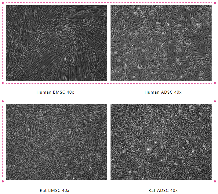
Morphological Assessment
Most MSCs exhibit a fibroblast-like morphology, growing on the surface of a substrate in a spindle-shaped or irregular triangular form. The cells contain an oval nucleus located in the center, with cytoplasmic extensions of varying lengths projecting outward. After several passages, MSCs show homogeneous, swirling arrangements. Nearly all MSCs are adherent, demonstrating strong attachment capability.
Surface Marker Identification
The identification of MSC surface markers is commonly performed using flow cytometry. MSCs belong to a heterogeneous cell population, and their surface antigens are not entirely specific. They can simultaneously express surface markers of mesenchymal, endothelial, and epithelial cells, such as adhesion molecules, growth factor receptors, cytokine receptors, and members of the integrin family.
The standard identification criteria for MSCs are as follows:
- Positive markers (≥90%) : CD105, CD73, CD90 (≥90% positive).
- Negative markers (≤5%) : CD45, CD34, CD14 or CD11b, CD79α or CD19, HLA‑DR.
- MSCs derived from different tissues may share common markers such as CD29 and CD44.
MSC Surface Marker Profile
| Marker | Expression | Threshold | Notes |
|---|---|---|---|
| CD105 (Endoglin) | Positive | ≥ 90% | TGF‑β co-receptor; classical MSC surface marker |
| CD73 (NT5E) | Positive | ≥ 90% | Ecto‑5′‑nucleotidase; MSC characteristic marker |
| CD90 (THY1) | Positive | ≥ 90% | Glycoprotein associated with MSC function |
| CD29 (ITGB1) | Positive | N/A | Frequently expressed in MSCs |
| CD44 | Positive | N/A | Hyaluronan receptor; commonly expressed MSC marker |
| CD45 | Negative | ≤ 5% | Pan‑leukocyte marker |
| CD34 | Negative | ≤ 5% | Hematopoietic/endothelial progenitor marker |
| CD14 or CD11b | Negative | ≤ 5% | Monocyte/macrophage/integrin markers |
| CD79α or CD19 | Negative | ≤ 5% | B‑cell lineage markers |
| HLA‑DR | Negative | ≤ 5% | MHC class II; typically absent in MSC |
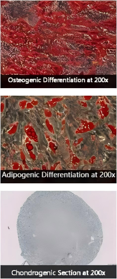
Multipotent Differentiation Potential
MSCs possess the ability to differentiate in vitro into osteoblasts, chondrocytes, adipocytes, hepatocytes, and other lineage-specific cells. Therefore, assessing their multipotent differentiation potential is also an important method of identification.
The commonly used staining methods are:
- Osteogenic differentiation: Alizarin Red staining
- Adipogenic differentiation: Oil Red O staining
- Chondrogenic differentiation: Alcian Blue staining or suspension culture
MSC Trilineage Differentiation
These three approaches—morphological assessment, surface marker identification, and multipotent differentiation potential—are standard and widely used methods for MSC identification. Have you learned them all?
Q & A
1. What does "three positive and five negative" mean in MSC identification?
It refers to the expression of CD73, CD90, and CD105 (positive) and the absence of CD45, CD34, CD14/CD11b, CD79α/CD19, and HLA‑DR (negative) within defined thresholds. This immunophenotype corresponds to the minimal criteria widely accepted for MSC identification. See ISCT position statement: Dominici et al., Cytotherapy, 2006; PubMed ID: 16923606.
2. What if CD105 positivity is below 90%?
Reduced CD105 expression can result from donor variability, tissue source, passage number, culture conditions, or gating strategy. MSC identity should be evaluated comprehensively using morphology, the full immunophenotypic panel, and trilineage differentiation. If CD105 is borderline, check the antibody clone, compensation, and gating strategy; repeating analysis after recovery in standard media may be necessary. Reference: ISCT minimal criteria.
3. Are CD31 (PECAM1) and CD11b mandatory negatives for MSC?
CD11b (ITGAM) is part of the negative marker set to exclude monocyte/macrophage contamination. CD31, an endothelial marker, is generally absent in MSC and its negativity helps exclude endothelial cells. See ISCT criteria: PubMed 16923606.
4. Which stains are used to verify MSC trilineage differentiation?
Osteogenesis: Alizarin Red S (calcium deposition).
Adipogenesis: Oil Red O (neutral lipids).
Chondrogenesis: Alcian Blue (glycosaminoglycans).
These stains provide qualitative confirmation of lineage‑specific matrix or lipid accumulation.
5. Are all adherent bone marrow cells MSC?
No. The initial adherent fraction contains a heterogeneous stromal population. MSC enrichment occurs over time through selective adherence and expansion, but identity must be confirmed via immunophenotyping and trilineage differentiation. Reference: Dominici et al., Cytotherapy 2006.
6. Do MSC express PDPN (podoplanin) ?
PDPN is not part of the classical MSC minimal criteria. Its expression varies depending on species and tissue source and is not required for MSC identification. Focus on the core panel (CD73, CD90, CD105) and required negative markers.
7. Can MSC identity be established by STR profiling alone?
No. Short tandem repeat (STR) profiling verifies sample identity (human source verification/contamination control) but does not determine cell phenotype. MSC identification requires morphology, immunophenotype, and functional differentiation.
8. What are common synonyms for CD73, CD90, and CD105?
CD73 = NT5E (ecto‑5′‑nucleotidase); CD90 = THY1; CD105 = Endoglin. These names may appear in product datasheets and literature.
9. What immunophenotype should rat or mouse MSC show by flow cytometry?
Rodent MSC generally follow the same pattern: CD73/CD90/CD105 positive and hematopoietic/endothelial markers negative (e.g., CD45, CD34, CD11b). Exact expression can vary with strain and tissue; confirm with lineage differentiation.
Related OriCell Products
- SD Rat Bone Marrow Mesenchymal Stem Cells
- F344 Rat Bone Marrow Mesenchymal Stem Cells
- Wistar Rat Bone Marrow Mesenchymal Stem Cells
- C57BL/6 Mouse Bone Marrow Mesenchymal Stem Cells
- Balb/c Mouse Bone Marrow Mesenchymal Stem Cells
- SD Rat Adipose‑derived Mesenchymal Stem Cells
- C57BL/6 Mouse Adipose‑derived Mesenchymal Stem Cells
- Complete Medium for Rat Bone Marrow MSC
- Complete Medium for Mouse Bone Marrow MSC
About Cyagen OriCell™
Cyagen OriCell™ is a Cyagen brand focused on the research and development of cell biology products, including stem cells, primary cells, and cell lines, as well as cell culture reagents and technical services. Serving universities, research institutes, hospitals, CROs, and CDMOs worldwide, Cyagen OriCell™ has accumulated extensive expertise in cell isolation and culture. The team has developed “spatial replication” culture technology to rapidly establish growth‑supportive environments, and runs an Antibiotic‑Free process grounded in strict environmental, materials, and personnel controls. Cyagen OriCell™ provides end‑to‑end solutions—from MSC isolation and identification to directed differentiation and assay services.
Cyagen OriCell™’s offerings are cited in over 10,000 publications, with a cumulative impact factor exceeding 90,000 and more than 160,000 citations, and the team has supported more than 3,000 research groups. Products are used by tens of thousands of customers across dozens of countries and regions.

