
Trilineage Differentiation and Identification of Mesenchymal Stem Cells (MSCs)
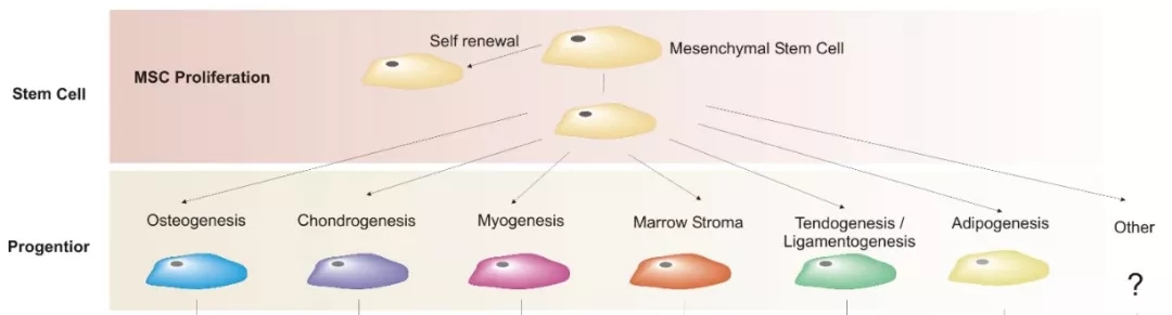
Mesenchymal stem cells (MSCs) are adult stem cells with self-renewal capability and multipotent differentiation potential. They originate from the immature embryonic connective tissue of the mesoderm during early embryogenesis. In 1968, German scientist Friedenstein first identified MSCs in bone marrow, after which they were successfully isolated from various tissues, including adipose tissue, umbilical cord, and dental pulp. As multipotent stem cells, MSCs can differentiate into multiple lineages. In 1999, Pittenger et al. first reported in Science the successful in vitro induction of bone marrow–derived MSCs into adipocytes, osteoblasts, and chondrocytes. Since then, MSCs have also been induced to differentiate into muscle cells, ligament tissue, hepatocytes, cardiomyocytes, endothelial cells, and other cell types.
At present, the identification of mesenchymal stem cells (MSCs) primarily relies on three aspects:
- morphological observation (e.g., fibroblast-like morphology, central oval nuclei, and whirlpool- or radial-like growth at high density);
- surface marker detection (such as CD29 and CD44);
- multilineage differentiation potential assessment.
The term "multilineage differentiation potential" usually refers to osteogenic, adipogenic, and chondrogenic differentiation, collectively known as the trilineage differentiation of MSCs. Although the induction procedures and identification methods for trilineage differentiation have become relatively well established, their underlying mechanisms may not yet be widely understood.
To facilitate a better understanding of MSC trilineage differentiation, this article provides a preliminary summary of the general differentiation processes, the characteristic markers that appear during differentiation, and the corresponding detection methods.
MSC Trilineage Differentiation: Principles and Assays
Osteogenesis (Bone Formation)
The differentiation of MSCs into osteoblasts serves as a powerful tool for understanding the mechanisms of bone formation and is also used to study hereditary disorders such as osteoporosis, bone repair defects, and osteogenesis imperfecta.
Like other tissues and organs, bone tissue undergoes continuous cellular metabolism, known as bone metabolism. This process can be broadly divided into two phases: bone formation and bone remodeling. Bone formation predominates during the developmental stage of an individual, whereas bone remodeling continues throughout the entire lifespan. Osteogenic differentiation is a key step in bone formation, during which bone marrow–derived MSCs undergo a complex multistage process—transitioning from osteoprogenitor cells to pre-osteoblasts, then to mature osteoblasts, and finally to terminally differentiated osteocytes. This process involves a network of intercellular and intracellular signaling events, including signaling pathways, transcription factors, growth factors, and microRNAs, which together form a complete negative feedback loop regulating bone metabolism. The transitions between different cell populations are gradual, representing a continuum of developmental stages.
The detailed process is as follows:
- Differentiation into Osteoprogenitor Cells. Under the stimulation of multiple cytokines, hormones, and other specific physicochemical factors, MSCs are induced to differentiate into osteoprogenitor cells.
- Proliferation of Pre-Osteoblasts. The osteoprogenitor cells then further differentiate into pre-osteoblasts and enter a rapid proliferation phase.
- Early Osteogenic Markers. As proliferative capacity decreases, genes associated with extracellular matrix (ECM) maturation—such as alkaline phosphatase, type I collagen, and matrix Gla protein—are activated. During this stage, osteoblasts synthesize and secrete organic matrix to form osteoid, mainly composed of type I collagen. The expression of alkaline phosphatase serves as an early marker of osteogenic differentiation and maturation.
- Osteoblast Maturation and Mineralization. Mature osteoblasts express ECM mineralization–related proteins, primarily osteocalcin and osteopontin, leading to a substantial increase in their mineralization activity.
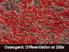
In vitro identification of osteogenesis
Corresponding to the in vivo process of osteogenic differentiation, in vitro identification of osteogenic differentiation is primarily performed by detecting biological markers of osteoblasts and their associated marker genes. Short-term osteogenic differentiation can be detected by alkaline phosphatase assays (ALP), while long-term osteogenic differentiation is identified by Alizarin Red S staining of mineralized bone nodules.
Specifically, the following three markers are detected:
- Alkaline Phosphatase (ALP): Following staining with BCIP/NBT substrate, alkaline phosphatase appears blue under the microscope, whereas undifferentiated cells show no obvious color. The osteogenic capacity can be evaluated and compared by quantifying the average expression intensity of alkaline phosphatase in cells across different samples. This can be achieved by comparing the percentage of osteogenic differentiated cells in each sample, measuring the average gray value of ALP-stained images (with fixed imaging parameters), or by dissolving the blue precipitate and measuring absorbance.
- Calcium Nodules: Prolonged osteogenic induction causes calcium ions to precipitate as calcium salts, forming "bone nodules". These bone nodules can be visualized through Alizarin Red S staining, which reacts with calcium to produce a deep red compound, staining the deposited calcium nodules bright red. The extent of osteogenic differentiation is indicated by the area and intensity of the staining.
- Osteoblast-Specific Marker Genes: Following osteogenic differentiation, MSCs express characteristic osteoblast genes such as ALP and Runx2 (runt-related transcription factor 2). RT-PCR can be used to compare osteogenic differentiation differences between experimental groups.
Osteogenic Induction of BMSCs
Adipogenesis (Fat Formation)
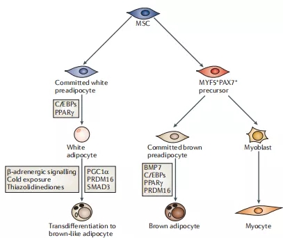
Adipose tissue is composed of adipocytes and plays a critical role in maintaining energy storage and metabolic homeostasis. There are two types of adipose tissue. White adipose tissue (WAT), mainly located in the subcutaneous and visceral depots, is associated in its visceral form with obesity, pathological conditions, and insulin resistance, whereas subcutaneous WAT can improve glucose tolerance. Brown adipose tissue (BAT), which is more discretely distributed along the paravertebral region, clavicles, and around the adrenal glands, functions primarily in thermogenesis. Despite their distinct locations and functions, both types of adipose tissue share similar differentiation characteristics and originate from mesenchymal stem cells.
Mesenchymal stem cell (MSC) adipogenic differentiation occurs in two stages. First, MSCs differentiate into preadipocytes; then, under specific cellular stimuli, such as C/EBP family members and PPAR-γ, they ultimately differentiate into mature adipocytes characterized by prominent lipid droplets.
In vitro identification of adipogenesis
Detection of lipid droplets in adipogenically induced cells. After Oil Red O staining, intracellular lipid droplets in differentiated adipocytes appear distinctly red under the microscope, whereas undifferentiated cells show no obvious coloration. The degree of adipogenic differentiation can be evaluated by quantifying the percentage of adipogenically differentiated cells in each sample.
Assessment of adipogenic marker gene expression. Following adipogenic differentiation, MSCs exhibit marked expression of adipocyte-specific genes such as LPL and PPARγ. RT-PCR can be used to measure the expression levels of these genes and to compare differences in adipogenic differentiation among different experimental groups.
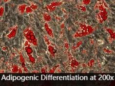
Adipogenic Induction of BMSCs
Chondrogenesis (Cartilage Formation)
Damage or loss of cartilage is a key pathological change in many orthopedic diseases, including osteoarthritis (OA). Due to the unique anatomical and structural characteristics of cartilage, such as the absence of blood vessels and lymphatic vessels, its intrinsic self-repair capacity is extremely limited, making artificial repair of cartilage defects of great clinical importance. In recent years, with the discovery and in-depth study of mesenchymal stem cells (MSCs), their wide range of tissue sources, strong proliferative capacity, multilineage differentiation potential, and good biocompatibility with various three-dimensional scaffold materials have made MSCs a highly promising cell source for cartilage repair.
During chondrogenic differentiation of MSCs, the cells first retract their processes and aggregate into clusters. Cells located in the center of these clusters then proliferate and differentiate into large, round chondroprogenitor cells. These chondroprogenitor cells produce extracellular matrix and fibers, mainly type II collagen. As the amount of matrix increases, the cells become surrounded in lacunae and further differentiate into mature chondrocytes.
In vitro identification of chondrogenesis
In vitro identification of chondrogenic differentiation mainly focuses on the cartilage-specific marker type II collagen. This includes detecting type II collagen expression by immunohistochemistry and Western blot, as well as assessing type II collagen mRNA expression by RT-PCR, thereby evaluating chondrogenesis from different perspectives. In addition, acidic glycosaminoglycans in the extracellular matrix can be visualized by Alcian Blue 8GX staining, and toluidine blue is commonly used to detect acidic mucopolysaccharides within chondrocytes.
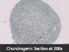
Chondrogenic Induction of BMSCs
Services and Reagents for MSC Differentiation
OriCell™ has extensive expertise in stem cell induction and differentiation, supported by a mature technology platform to perform a wide range of MSC differentiation potential assays.
Below are representative induction media and assay kits:
- Osteogenic Differentiation Medium (Human MSCs)
- Adipogenic Differentiation Medium (Human MSCs)
- Chondrogenic Differentiation Medium (Human MSCs)
- MSC Characterization Kit (Human)
- MSC Characterization Kit (Rat)
- MSC Characterization Kit (Mouse)
Common reagents used in MSC culture and staining include:
- Alizarin Red S Solution
- Oil Red O Solution
- Alcian Blue 8GX Solution
- L‑Glutamine
- NEAA Supplement
- ITS Supplement
- DMEM High Glucose Complete Medium
References
Cristancho AG, Lazar MA. Forming functional fat: a growing understanding of adipocyte differentiation. Nat Rev Mol Cell Biol. 2011;12(11):722–734. doi:10.1038/nrm3198.
About OriCell™
OriCell™ is Cyagen's sub‑brand dedicated to stem cell reagents and technologies. As a STEM cell supplier with 19 years of history, OriCell™ has developed hundreds of stem cell products. Our primary cells, fetal bovine serum, cell culture media and cryopreservation media have served tens of thousands of customers and contributed to over 13,524 papers—including CNS (Cell, Nature, Science). For inquiries, please contact us.
Q & A
1. What is MSC trilineage differentiation?
It refers to in vitro induction of MSCs into osteogenic, adipogenic, and chondrogenic lineages followed by identification using lineage‑specific markers and assays.
2. What methods are used to identify osteogenic differentiation ?
Short‑term assays assess ALP activity; long‑term assays detect calcified bone nodules with Alizarin Red S. Marker genes such as ALP and Runx2 are evaluated by RT‑PCR.
3. What methods are used to identify adipogenic differentiation?
Oil Red O staining visualizes neutral lipid droplets in adipocytes; PPARγ and LPL expression is measured by RT‑PCR. See Oil Red O Solution.
4. What methods are used to identify chondrogenic differentiation?
Type II collagen is assessed by immunohistochemistry/Western blot/RT‑PCR (COL2A1). Acidic glycosaminoglycans in the extracellular matrix are detected by Alcian Blue 8GX staining.
5. What surface markers are commonly used to characterize MSCs?
Common markers include CD29 and CD44; characterization can be performed using MSC Characterization Kits.
6. What factors determine the outcomes of differentiation?
Induction conditions, stimulatory factors, and assay timing influence outcomes; consistent imaging and quantification improve comparability across groups.
7. What induction media are recommended for MSC trilineage assays?
For human MSCs: osteogenic, adipogenic, and chondrogenic media.
8. How can osteogenic and adipogenic differentiation be quantified?
By percentage of positive cells, staining intensity/area, and marker gene expression measured under fixed parameters.
9. What function does type II collagen serve in chondrogenesis?
Type II collagen is a signature ECM component of cartilage; its detection confirms chondrogenic matrix formation.
10. Where are MSC products and services available?
Visit OriCell™ for stem cells, characterization kits, induction media, and staining solutions.
About Cyagen OriCell™
Cyagen OriCell™ is a Cyagen brand focused on the research and development of cell biology products, including stem cells, primary cells, and cell lines, as well as cell culture reagents and technical services. Serving universities, research institutes, hospitals, CROs, and CDMOs worldwide, Cyagen OriCell™ has accumulated extensive expertise in cell isolation and culture. The team has developed “spatial replication” culture technology to rapidly establish growth‑supportive environments, and runs an Antibiotic‑Free process grounded in strict environmental, materials, and personnel controls. Cyagen OriCell™ provides end‑to‑end solutions—from MSC isolation and identification to directed differentiation and assay services.
Cyagen OriCell™’s offerings are cited in over 10,000 publications, with a cumulative impact factor exceeding 90,000 and more than 160,000 citations, and the team has supported more than 3,000 research groups. Products are used by tens of thousands of customers across dozens of countries and regions.

