
What Are Fibroblasts? A Guide to Understanding and Isolating Human Dermal Fibroblasts (HDFs)
Did you know that the skin is the largest organ of the human body? It serves as the primary barrier against the external environment and plays essential roles in thermoregulation, sensation, self-renewal, and immune defense. Within the collagen-rich dermis lies the focus of this guide: the dermal fibroblast. In this guide, we outline HDF functions, marker profiles, and a practical isolation protocol. Related reagents include Complete Medium for Human Fibroblasts, Phosphate-Buffered Saline (PBS), and 0.1% Collagenase Type I.
Human Dermal Fibroblasts (HDFs): Role and Marker Profile
Human Dermal Fibroblasts (HDFs) are the principal stromal cells of the dermis, responsible for synthesizing the extracellular matrix (ECM) and collagen, thereby maintaining structural integrity. Although fibroblasts are not classified as mesenchymal stem cells (MSCs), they share several MSC-like characteristics. They express similar surface markers—positive for CD105, CD73, and CD90, and negative for CD45, CD34, CD14, CD11b, CD79a, CD19, and HLA-DR—and can be induced in vitro to differentiate into adipocytes, osteoblasts, and chondrocytes.
However, another perspective suggests that fibroblasts are highly heterogeneous, and not all fibroblast populations display MSC-like properties. Therefore, they should not be categorized as true mesenchymal stem cells. Despite the ongoing debate regarding their identity, fibroblasts exhibit remarkable functional versatility and remain indispensable model cells for studies on skin aging mechanisms and wound healing.
Key Features
- Location: Dermis, within collagen-rich connective tissue
- Functions: ECM and collagen synthesis, structural support, and wound repair
- Applications: Skin aging research and injury repair modeling
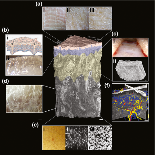
Skin Structure Showing the Dermis and the Location of Dermal Fibroblasts
From Theory to Practice: Isolating Primary HDFs
The following is a standardized protocol for isolating human dermal fibroblasts (HDFs) using enzymatic digestion.
1. Materials and Reagents Preparation
- Sample: Human skin tissue containing dermis, ≥5 mm², collected within 12 hours. Preserve in basal medium supplemented with 1% penicillin-streptomycin (Pen-Strep) at 4°C.
- Reagents: OriCell™ PBS (1X), OriCell™ Complete Medium for Human Fibroblasts, OriCell™ Collagenase Type I (0.1%).
- Consumables: 50 mL collection bottles; 50 mL tubes (5–10); 10 mL pipettes (4); T25/T75 flasks.
- Instruments: Hemostats, ophthalmic forceps, ophthalmic scissors.
2. Isolation Protocol (Step-by-Step)
Step 1: Washing and Disinfection
- Transfer the skin tissue from the collection bottle into a 50 mL centrifuge tube containing 20 mL PBS with antibiotics (Pen-Strep). Rinse repeatedly for 3 minutes to completely remove blood and debris.
- Transfer the tissue into another 50 mL tube containing 20 mL of 75% ethanol for surface sterilization. Incubate for 40 seconds, then rinse twice with PBS.
Note: Skin tissue is exposed to the external environment and is non-sterile. Maximize washing and disinfection before digestion.
Step 2: First Digestion (Pre-Digestion)
- Add an equal volume of freshly prepared, pre-warmed (37 °C, 30 min in CO₂ incubator) Collagenase Type I solution.
- Seal the tube with parafilm, shake vigorously for 5–10 seconds, and place it at 4 °C for overnight digestion.
- The next day, remove the epidermis and adipose tissue completely, then finely mince the remaining dermal tissue.
Note: Fibroblasts reside in the dermis. Remove all tissue outside the dermis to minimize contamination.
Step 3: Second Digestion
- Add an equal volume of freshly prepared, pre-warmed Collagenase Type I solution.
- Seal with parafilm, shake for 5–10 seconds, and incubate in a shaking water bath at 37 °C, 150 rpm for 90 minutes.
- Terminate digestion by adding 2 mL complete medium.
- Adjust the total volume to 90 mL with PBS and divide into two 50 mL centrifuge tubes.
- Centrifuge at 250 × g for 6 minutes.
Step 4: Washing and Filtration
- Discard the supernatant carefully (do not pour directly).
- Resuspend the pellet in fresh medium, then combine into one tube.
- Adjust the volume to 45 mL with PBS and centrifuge again at 250 × g for 6 minutes.
- Resuspend the pellet in 10 mL PBS, pipette gently to disperse cells, and pass through a 100 µm cell strainer.
- Divide the filtrate into two 50 mL tubes, add PBS to a final volume of 30 mL.
- Collect small tissue fragments from the strainer. Evenly spread these fragments on the bottom of T25 flasks designated for seeding.
Step 5: Cell Collection and Seeding
- Centrifuge at room temperature, 250 × g for 6 minutes, and carefully remove the supernatant.
- Wash the cell pellet with 10 mL PBS, centrifuge again (250 × g, 6 minutes), discard the supernatant, and resuspend in 3 mL complete culture medium.
- Seed the cell suspension into one T25 flask and incubate at 37 °C with 5% CO₂ under standard culture conditions.
- Inspect after 24 hours for attachment and signs of contamination.
Medium Change (Half-Change Method)
- Perform the first half-medium change on day 3 after initial seeding.
- Transfer the medium from the culture flask into a 50 mL centrifuge tube and centrifuge at 250 × g for 6 minutes.
- After centrifugation, add 1.5 mL of the recovered (old) medium and 1.5 mL of fresh complete medium to the T25 flask to complete the half-medium change.
- Place the culture flask in a CO₂ incubator maintained at 37 °C, 5% CO₂, and saturated humidity.
- After the first change, perform half-medium replacement every 3 days (adjust as needed based on cell growth).
- Continue half-medium changes until the culture reaches approximately 90% confluence, at which point subculture is required.
Passaging (Subculture)
Protocol (P0 → P1 and Subsequent)
- Prewarm complete medium, PBS, and trypsin to 37 °C.
- Aspirate the culture medium from the vessel. Wash the cells twice with PBS (approximately 3 mL for a T25 flask, 6 mL for a T75 flask). Handle gently to ensure thorough but careful washing. Then remove the PBS completely.
- Add trypsin (approximately 1.5 mL for a T25 flask, 3 mL for a T75 flask) and distribute evenly to ensure full contact with the cell surface.
- Observe cell detachment under a microscope. When 70%–80% of cells have shrunk and become rounded, gently tap the side of the flask to detach them.
- Immediately add complete medium (approximately 3 mL for a T25 flask, 6 mL for a T75 flask) to neutralize trypsin. Gently swirl the flask to mix.
- Using a pipette, aspirate and gently pipette the cell suspension several times along the bottom of the culture vessel to dislodge any remaining adherent cells.
- Transfer the cell suspension into a centrifuge tube. Rinse the culture vessel once with PBS (3 mL for a T25 flask, 6 mL for a T75 flask) to collect remaining cells, and combine all suspensions.
- Centrifuge at 250 × g for 4 minutes.
- Carefully remove the supernatant, then resuspend the pellet in 2 mL of complete medium. Gently pipette to fully disperse and homogenize the cells.
- Seed cells at a density of (2.5 – 4) × 10⁴ viable cells/cm² into appropriate culture vessels.
- Gently swirl the flask to distribute the cells evenly and place it in a CO₂ incubator at 37 °C, 5% CO₂, and saturated humidity.
- Observe cell morphology on the following day. If numerous floating cells are present, perform a medium change.
- Once cells reach approximately 90% confluence, proceed to subculture or cryopreservation.
Note:
- Avoid vigorous pipetting and bubbles to prevent damage and loss of cells.
- Cell density is critical for successful fibroblast culture. When possible, perform manual cell counting to ensure accurate seeding density. If precise counting is not available, passage based on appropriate dilution ratios. Typically, human dermal fibroblasts are subcultured at a 1:3 ratio and reach confluence suitable for passaging within 72 hours. Adjust as needed according to growth conditions.
- Under normal conditions, HDFs reach confluence within 72 hours and do not require medium exchange during this period. Frequent medium changes can disrupt the microenvironment necessary for stable cell growth.
Related Q&A
1. What are human dermal fibroblasts (HDFs)?
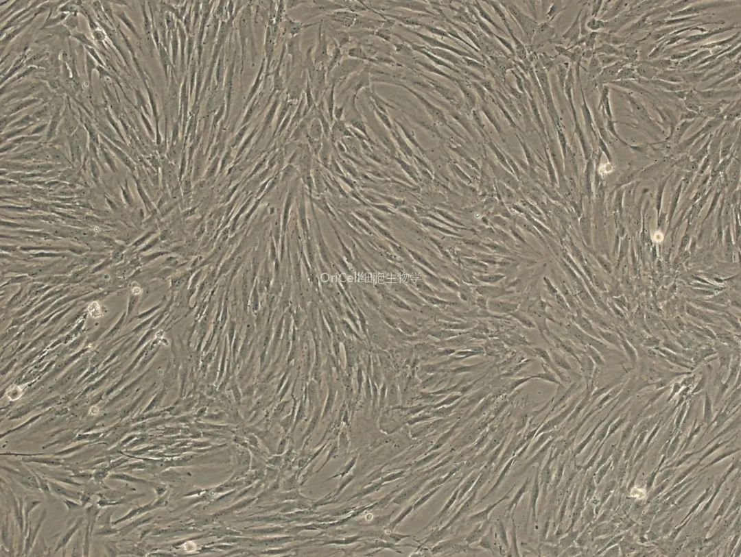
Human dermal fibroblasts are the primary stromal cells of the dermis. They synthesize collagen and extracellular matrix (ECM), support tissue repair, and typically display elongated, spindle-shaped morphology.
2. Are fibroblasts classified as stem cells?
No. Fibroblasts are not classified as mesenchymal stem cells (MSCs). While some subpopulations show MSC-like properties, fibroblast heterogeneity prevents them from being universally defined as MSCs.
3. Do dermal fibroblasts express CD34 or CD45?
Generally, HDFs are CD34⁻/CD45⁻. Marker expression can vary depending on tissue origin, donor age, and cellular state.
4. What is the common abbreviation for fibroblasts?
Fibroblasts are commonly abbreviated as FBs or HDFs (human dermal fibroblasts) when referring specifically to dermal origin.
5. What medium is recommended for culturing HDFs?
Use a complete fibroblast medium formulated for dermal fibroblasts. (Example: Complete Medium for Human Fibroblasts.)
6. Can HDFs undergo differentiation?
Yes. Under specific induction conditions, fibroblasts can exhibit MSC-like differentiation potential, differentiating into adipogenic, osteogenic, or chondrogenic lineages. However, these capabilities depend on the context and are not universally observed.
7. Are dermal fibroblasts positive for CD90?
Many HDFs express CD90 (Thy-1), as well as CD73 and CD105. However, marker expression can vary among donors and fibroblast subsets.
8. Do HDFs require frequent medium changes?
No. Most cultures require medium changes every ~72 hours. Over-frequent changes may disrupt the microenvironment and slow attachment or proliferation.
9. How can fibroblast identity be confirmed without immunofluorescence?
Evaluate characteristic spindle-shaped morphology, adherence properties, growth behavior, and absence of hematopoietic markers. Additional functional assays (e.g., collagen production) can provide further confirmation.
10. From which tissues can fibroblasts be isolated?
Dermis is the most common source. Fibroblasts can also be isolated from various connective tissues and organs. However, isolation protocols differ depending on tissue structure.
About Cyagen OriCell™
Cyagen OriCell™ is a Cyagen brand focused on the research and development of cell biology products, including stem cells, primary cells, and cell lines, as well as cell culture reagents and technical services. Serving universities, research institutes, hospitals, CROs, and CDMOs worldwide, Cyagen OriCell™ has accumulated extensive expertise in cell isolation and culture. The team has developed “spatial replication” culture technology to rapidly establish growth‑supportive environments, and runs an Antibiotic‑Free process grounded in strict environmental, materials, and personnel controls. Cyagen OriCell™ provides end‑to‑end solutions—from MSC isolation and identification to directed differentiation and assay services.
Cyagen OriCell™’s offerings are cited in over 10,000 publications, with a cumulative impact factor exceeding 90,000 and more than 160,000 citations, and the team has supported more than 3,000 research groups. Products are used by tens of thousands of customers across dozens of countries and regions.

