
3T3-L1 Cell Line: A Robust and Classic Model for Adipose Metabolism
What Is the 3T3-L1 Cell Line?
The 3T3-L1 mouse embryo fibroblast (preadipocyte) cell line is a continuous subclone derived from 3T3 (Swiss mouse) by clonal isolation. When 3T3-L1 cells transition from rapid division to confluence and contact inhibition, they convert from a preadipocyte state toward adipocyte-like cells. In culture, high serum content can promote intracellular lipid accumulation in 3T3-L1 cells.
3T3-L1 Cell Line Overview
Culture Conditions (37°C, 5% CO2)
| Tissue origin | Mouse embryo |
| Cell characteristics | Fibroblast-like; adherent growth |
| Culture conditions | 95% air; 5% CO2; 37°C |
| Culture medium | DMEM + 10% FBS + 1% Glu + 1% NEAA + 1‰ SP |
| Doubling time | 38–46 h |

Applications of the 3T3-L1 Cell Line
3T3-L1 cells not only can be stably passaged but also exhibit strong specificity for differentiation into adipocytes, making them an internationally recognized in vitro model for studying adipose metabolism. They have high research value for investigating adipocyte proliferation and differentiation. Below are two example applications of the 3T3-L1 cell line.
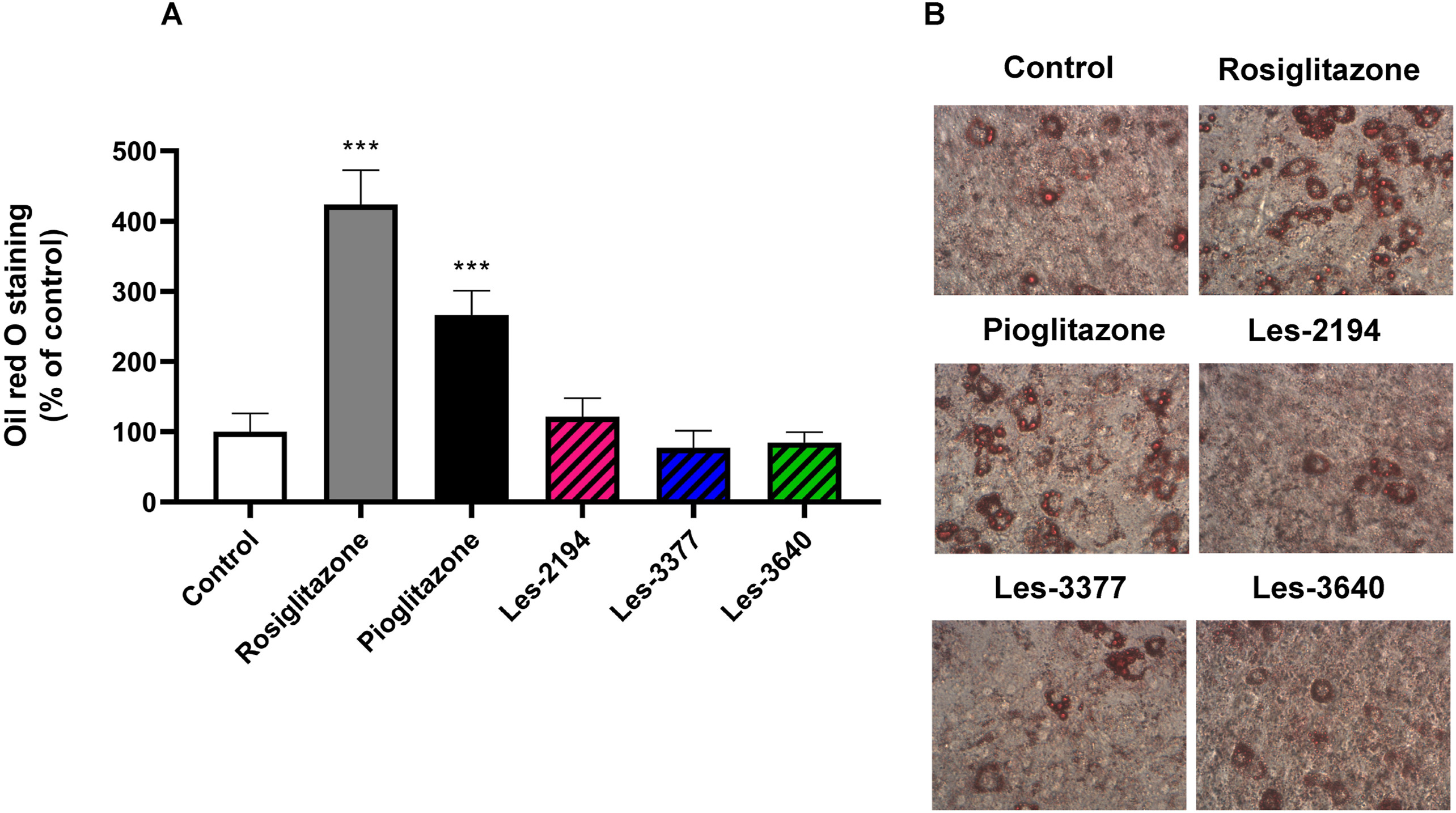
Oil Red O Staining After 14 Days of Differentiation
4‑Thiazolidinone-based derivatives do not affect 3T3-L1 differentiation into adipocytes [1]
Thiazolidinediones (TZDs) are widely used to reduce insulin resistance in type 2 diabetes, and are thought to act primarily through activation of the PPARγ receptor. Given that rosiglitazone, pioglitazone, Les-2194, Les-3377, and Les-3640 share structural similarity with TZDs, their effects on 3T3-L1 cell-line differentiation into adipocytes were examined to probe the potential mechanisms of TZDs in adipocyte differentiation. At 2 μM, these compounds did not affect 3T3-L1 cell viability nor trigger apoptosis. After 14 days of induction, Oil Red O staining showed that rosiglitazone and pioglitazone increased lipid accumulation in 3T3-L1 cells (by 323.49% and 166.13% versus control, respectively), a hallmark of mature adipocytes. To confirm full differentiation, rosiglitazone was also found to increase expression of PPARγ and NF-κB-specific proteins.
PPARγ and NF‑κB Expression During Adipocyte Differentiation
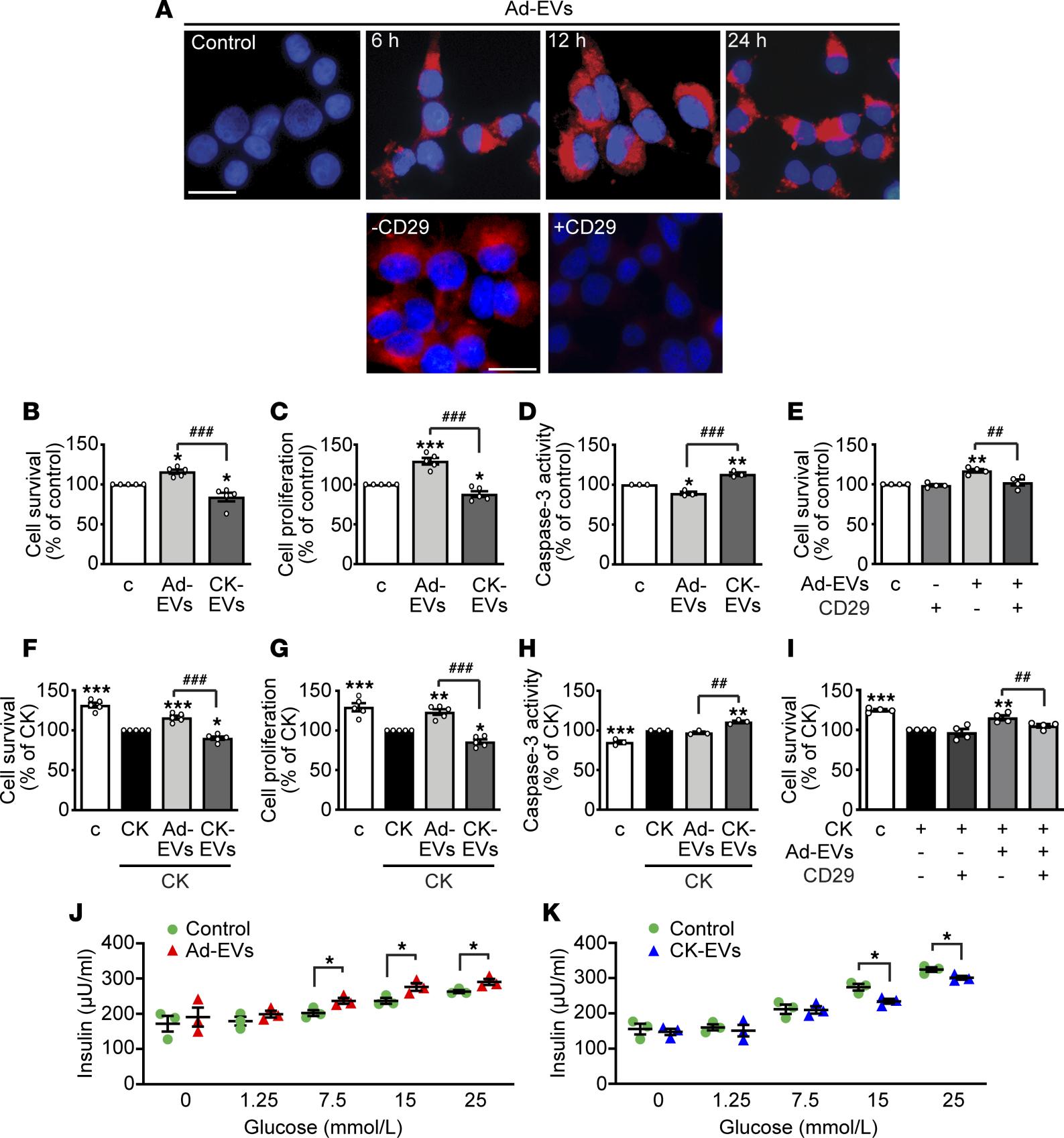
Adipocyte-derived extracellular vesicles regulate survival and function of pancreatic β cells [2]
Loss of pancreatic β cells is closely associated with type 1 diabetes and late-stage type 2 diabetes. As critical intercellular messengers, extracellular vesicles mediate communication between cell types. Studies show that extracellular vesicles derived from 3T3-L1 adipocytes exert both positive and negative effects on the survival, proliferation, and function of β cells and human islets in vitro, depending on adipocyte state and adipose-tissue origin. In vivo, healthy adipose tissue tends to have beneficial effects, whereas under pathological conditions such as obesity and insulin resistance, crosstalk between adipocytes and β cells may form a negative feedback loop, further amplifying insulin resistance and β-cell dysfunction. Understanding the mechanisms of extracellular vesicles may aid the prevention of β-cell loss and inform new strategies for diabetes treatment.
References
Szychowski KA, Skóra B, Kryshchyshyn-Dylevych A, Kaminskyy D, Tobiasz J, Lesyk RB, Gmiński J. 4-Thiazolidinone-based derivatives do not affect differentiation of mouse embryo fibroblasts (3T3-L1 cell line) into adipocytes. Chem Biol Interact. 2021 Aug 25;345:109538. doi: 10.1016/j.cbi.2021.109538. Epub 2021 Jun 9. PMID: 34097888.
Gesmundo I, Pardini B, Gargantini E, Gamba G, Birolo G, Fanciulli A, Banfi D, Congiusta N, Favaro E, Deregibus MC, Togliatto G, Zocaro G, Brizzi MF, Luque RM, Castaño JP, Bocchiotti MA, Arolfo S, Bruno S, Nano R, Morino M, Piemonti L, Ong H, Matullo G, Falcón-Pérez JM, Ghigo E, Camussi G, Granata R. Adipocyte-derived extracellular vesicles regulate survival and function of pancreatic β cells. JCI Insight. 2021 Mar 8;6(5):e141962. doi: 10.1172/jci.insight.141962. PMID: 33539327; PMCID: PMC8021102.
Cell Culture and Product Information
In vitro cell culture is an important tool to model human development and disease. OriCell® maintains a large, comprehensive research cell bank and provides multiple stem cells, cell lines, and matched culture systems, such as 3T3-L1 cells and complete media, as well as adipogenic differentiation media. For inquiries, please contact us.
Complete Medium Composition (DMEM + 10% FBS)
| Product name | Catalog No. | Specification |
|---|---|---|
| OriCell™ 3T3-L1 Mouse Embryonic Fibroblast (Pre-Adipocyte) Cell Line | M5-0101 | 1×10^6 cells |
| OriCell™ Complete Medium For 3T3-L1 Cell Line | CMM5-0101 | 500 mL |
| Adipogenic Differentiation Medium For Mouse 3T3-L1 Cells | / | 200 + 100 mL |
Q&A (Long-Tail Keywords)
Q1. What is the 3T3-L1 cell line?
3T3-L1 is a mouse embryo fibroblast-derived preadipocyte line widely used as an in vitro model of adipogenesis and adipose metabolism.
Q2. What is the tissue origin of 3T3-L1 cells?
They originate from mouse embryo fibroblasts (derived by clonal isolation from the 3T3 line).
Q3. What are standard culture conditions for 3T3-L1 cells?
Typical conditions are 37°C, 5% CO2, and adherent growth in DMEM supplemented with 10% FBS and additives as listed in the table.
Q4. What is the typical doubling time of 3T3-L1 cells?
Approximately 38–46 hours under standard culture conditions.
Q5. What does Oil Red O staining indicate in 3T3-L1 cells?
Oil Red O staining detects neutral lipid accumulation, which increases after successful adipogenic differentiation.
Q6. Which compounds are often studied with 3T3-L1 to probe adipogenesis?
Thiazolidinediones (e.g., rosiglitazone and pioglitazone) are commonly examined for their effects on differentiation and lipid accumulation.
Q7. What is PPARγ’s role during 3T3-L1 adipocyte differentiation?
PPARγ expression increases during adipogenesis and is associated with mature adipocyte markers.
Q8. Are 3T3-L1 cells adherent or suspension?
They are fibroblast-like and grow adherently.
Q9. What medium formulation is commonly used for 3T3-L1 culture?
DMEM + 10% FBS with supplements such as 1% Glu, 1% NEAA, and 1‰ SP, as specified in the overview table.
Q10. How do adipocyte-derived extracellular vesicles affect pancreatic β cells?
Their effects can be beneficial or detrimental depending on adipocyte state and tissue origin; understanding these mechanisms informs diabetes research.
About Cyagen OriCell™
Cyagen OriCell™ is a Cyagen brand focused on the research and development of cell biology products, including stem cells, primary cells, and cell lines, as well as cell culture reagents and technical services. Serving universities, research institutes, hospitals, CROs, and CDMOs worldwide, Cyagen OriCell™ has accumulated extensive expertise in cell isolation and culture. The team has developed “spatial replication” culture technology to rapidly establish growth‑supportive environments, and runs an Antibiotic‑Free process grounded in strict environmental, materials, and personnel controls. Cyagen OriCell™ provides end‑to‑end solutions—from MSC isolation and identification to directed differentiation and assay services.
Cyagen OriCell™’s offerings are cited in over 10,000 publications, with a cumulative impact factor exceeding 90,000 and more than 160,000 citations, and the team has supported more than 3,000 research groups. Products are used by tens of thousands of customers across dozens of countries and regions.

