
How to Achieve Stunning Alizarin Red S Staining in Osteogenic Differentiation
This guide shows how to achieve consistent and reliable Alizarin Red S (ARS) staining for bright, easily visualized mineralized nodules during osteogenic differentiation. ARS is an anthraquinone derivative that chelates calcium salts, forming an orange‑red complex. For optimal results, we recommend pairing your workflow with OriCell™ Osteogenic Differentiation Media and OriCell™ Alizarin Red S Solution (Cat. No.: ALIR‑10001).
Overview: Principle & Use
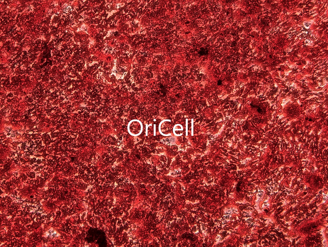
When mesenchymal stem cells (MSCs) are cultured in osteogenic media, they deposit calcium-rich minerals, forming distinct calcified nodules. ARS staining clearly visualizes these mineral deposits under brightfield microscopy. This enables qualitative assessment and, if desired, semi-quantitative analysis.
When to Perform ARS Staining
- Perform ARS staining only after calcified nodules are clearly visible under the microscope, rather than relying solely on culture duration.
- The optimal induction period may vary depending on the cell type and medium used. If calcified nodules are sparse, extend the induction period rather than staining prematurely.
Materials
- 1× PBS
- 4% Paraformaldehyde (PFA) Solution (or 10% Formalin)
- Alizarin Red S Solution
- Six-well plates
- Pipettes
- Microscope
Step-by-Step ARS Staining Protocol (6-Well Plate)
1. Wash Cells.
After completing osteogenic induction, carefully aspirate the osteogenic differentiation medium from the 6-well plate. Gently wash the cells 2–3 times with 1× PBS.
2. Fixation.
Add 2 mL of 4% paraformaldehyde (PFA) solution or 10% formalin to each well and fix the cells at room temperature for 30 minutes.
3. Wash after Fixation.
Aspirate the fixative and gently wash the cells 2–3 times with 1× PBS to completely remove residual fixative.
4. Staining.
Add 2 mL of Alizarin Red S working solution to each well and incubate at room temperature for 5–10 minutes.
5. Wash after Staining.
Aspirate the staining solution and gently wash 2–3 times with 1× PBS to remove excess stain.
6. Observation.
Add 2 mL of 1× PBS to each well and examine the mineralized nodules under a microscope.
7. Storage.
Seal the stained 6-well plate with parafilm and store at 4 °C. The plate can be kept for up to 2 weeks.
Practical Tips
- Handle the cells gently to prevent detachment of fragile mineralized nodules.
- Bring the ARS solution to room temperature before use.
- Adjust staining duration according to nodule visibility, not induction time.
- If staining appears weak, extend the incubation by 1–3 minutes, but avoid over-staining.
- Protect all solutions from light during storage and follow the kit instructions regarding pH and storage conditions.
Optional Quantification (CPC Method)
- After imaging, add 10% cetylpyridinium chloride (CPC) to each well to elute the bound ARS stain for 20–30 minutes.
- Measure the absorbance at 562 nm using a microplate reader.
- Normalize the results to cell number or total protein content, and include appropriate controls and replicates for reliable quantification.
Recommended Products
For robust osteogenic differentiation, we recommend our OriCell™ Osteogenic Differentiation Media tailored to your specific cell type. Each kit includes basal medium, premium fetal bovine serum, osteogenic supplements, and Alizarin Red S (ARS) solution. The Alizarin Red S Solution (Cat. No.: ALIR‑10001) is also available separately or as part of the osteogenic kit.
Popular Products
FAQs: Alizarin Red S Staining
1. What is the principle of ARS staining?
ARS chelates calcium ions, forming an orange-red complex that visualizes mineralized nodules in osteogenic cultures.
2. How long should I stain with ARS?
Typically 5–10 minutes at room temperature. Adjust the duration based on the visibility of nodules; extend slightly if staining is weak.
3. Can ARS solution be reused?
No. Do not reuse the working solution to prevent contamination and variability. Always prepare fresh solution according to the protocol.
4. Does ARS staining require a specific pH?
Use the kit-provided solution and store it in the dark. Avoid adjusting the pH unless the protocol explicitly specifies.
5. How do I quantify ARS staining?
Elute the bound dye with 10% cetylpyridinium chloride (CPC) and measure absorbance at 562 nm. For robust analysis, combine with image-based quantification (e.g., ImageJ).
6. ARS vs. Von Kossa: what's the difference?
ARS directly detects calcium via chelation, while Von Kossa indirectly detects phosphate using a silver nitrate reaction.
7. My plate appears too red — how to fix?
Reduce staining time, increase PBS washes, and ensure nodules are visible before staining. Avoid excessively long fixation.
8. Can I perform ARS in 24-well plates?
Yes. Scale reagent volumes proportionally (e.g., ~500 µL per well) based on surface area.
9. Why do nodules detach during processing?
Excessive handling or insufficient fixation can cause detachment. Use gentle pipetting and avoid aggressive washes.
*For research use only. Not for diagnostic or therapeutic applications.
About Cyagen OriCell™
Cyagen OriCell™ is a Cyagen brand focused on the research and development of cell biology products, including stem cells, primary cells, and cell lines, as well as cell culture reagents and technical services. Serving universities, research institutes, hospitals, CROs, and CDMOs worldwide, Cyagen OriCell™ has accumulated extensive expertise in cell isolation and culture. The team has developed “spatial replication” culture technology to rapidly establish growth‑supportive environments, and runs an Antibiotic‑Free process grounded in strict environmental, materials, and personnel controls. Cyagen OriCell™ provides end‑to‑end solutions—from MSC isolation and identification to directed differentiation and assay services.
Cyagen OriCell™’s offerings are cited in over 10,000 publications, with a cumulative impact factor exceeding 90,000 and more than 160,000 citations, and the team has supported more than 3,000 research groups. Products are used by tens of thousands of customers across dozens of countries and regions.

