
 EN
EN
A Step-by-Step Beginner Guide to Osteogenic, Adipogenic, and Chondrogenic Differentiation of Rat Tendon Stem Cells
At OriCell, we often receive questions from researchers working with rat tendon stem cells (TSCs), especially regarding how to successfully perform multi-lineage differentiation experiments and evaluate differentiation outcomes. Similar questions also appear frequently in Reddit discussions and lab forums, where researchers compare induction workflows, staining results, and troubleshooting tips for stem cell differentiation assays.
As a unique stem cell population, rat tendon stem cells have attracted increasing attention due to their important role in tendon regeneration and their potential for tissue engineering and regenerative medicine research. Their ability to differentiate into multiple cell lineages makes them a valuable model for studying tendon-related diseases, repair mechanisms, and cell-based therapeutic strategies.
To help researchers better understand the complete experimental workflow, OriCell scientists recently hosted an online seminar titled “The Generation Journey of Rat Tendon Stem Cells: From Tissue Collection to Characterization.” Following the positive feedback and questions received from researchers, our technical team prepared a detailed visual guide covering the complete process of osteogenic, adipogenic, and chondrogenic differentiation of tendon stem cells.
In this guide, our scientists will walk you through the key steps of tendon stem cell differentiation, including experimental preparation, induction conditions, and evaluation methods, helping researchers better understand the critical factors affecting successful differentiation.
Experimental Materials
- Primary cells: OriCell SD Rat Tendon Stem Cells (Cat. No. RASTA-01001)
- Culture medium: OriCell Complete Medium For Rat Tendon Stem Cells (Cat. No. RAXTA-90011)
- Induction kits:
OriCell Osteogenic Differentiation Medium For Rat Tendon Stem Cells (Cat. No. RASTA-90021)
OriCell Adipogenic Differentiation Medium For Rat Tendon Stem Cells (Cat. No. RASTA-90031)
OriCell Chondrogenic Differentiation Medium For Rat Tendon Stem Cells (Cat. No. RASTA-90041)
Osteogenic Differentiation
Core Steps for Osteogenic Differentiation of Tendon Stem Cells
- • Cell coating: Coat the culture vessel with 0.1% gelatin or poly-L-lysine solution, and incubate at 37°C for 30 min to enhance cell attachment.
- • Cell seeding: Prepare a single-cell suspension using OriCell SD Rat Tendon Stem Cells in the logarithmic growth phase. Seed the cells into the coated culture vessel at a density of 2 × 10⁴ cells/cm². During culture, use OriCell Complete Medium For Rat Tendon Stem Cells to maintain cell growth and proliferation.
- • Induction initiation: When the cells reach approximately 70% confluence, start induction. Aspirate the original culture medium and replace it with OriCell Osteogenic Differentiation Medium For Rat Tendon Stem Cells for osteogenic induction. Change the medium every 3 days. For preparation of the kit and detailed induction steps, please refer to the product manual.
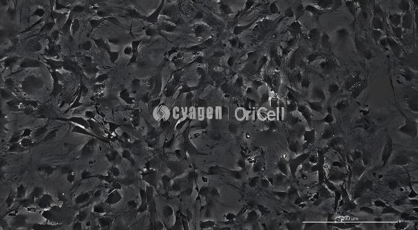
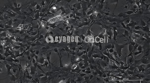
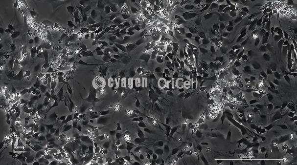
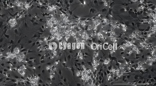
Images showing the induction process: From top to bottom: day 3, day 7, day 13, and day 19 of induction.
Staining and Identification
Alizarin Red staining is used to detect red calcium nodules and verify mineralization capacity.
Alizarin Red Staining Procedure
- Termination of induction and washing: Aspirate the osteogenic induction medium, and gently wash the cells 2–3 times with pre-cooled 1× PBS to remove residual medium.
- Cell fixation: Add fixative to each well and fix at room temperature for 30 min.
- Staining reaction: Aspirate the fixative. After washing twice with 1× PBS, add Alizarin Red working solution to cover the cells and stain at room temperature for 5–10 min.
- Termination of staining and washing: Aspirate the staining solution. Wash 2–3 times with 1× PBS until no floating background color remains, then add 1× PBS to keep the cells moist.
Staining result: Dense red particles indicate a high degree of mineralization and successful osteogenic differentiation. In contrast, no staining or weak staining indicates unsuccessful differentiation.
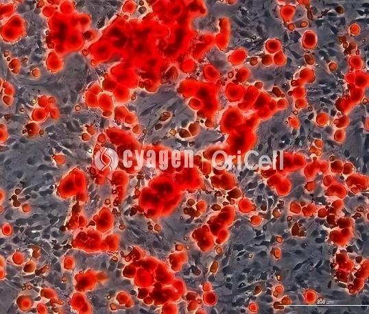
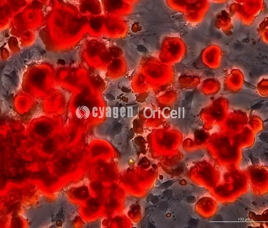
Alizarin Red staining result on day 19
Adipogenic Differentiation
Core Steps for Adipogenic Differentiation of Tendon Stem Cells
- • Cell coating: Coat the culture vessel with 0.1% gelatin or poly-L-lysine solution, and incubate at 37°C for 30 min to enhance cell attachment.
- • Cell seeding: Prepare a single-cell suspension using OriCell SD Rat Tendon Stem Cells in the logarithmic growth phase. Seed the cells into the coated culture vessel at a density of 3 × 10⁴ cells/cm². During culture, use OriCell Complete Medium For Rat Tendon Stem Cells to maintain cell growth and proliferation.
- • Induction initiation: When the cells reach 100% confluence, start induction. Aspirate the original culture medium and replace it with OriCell Adipogenic Differentiation Medium For Rat Tendon Stem Cells for adipogenic induction. In the early stage, perform cyclic induction using “3 days in Solution A followed by 1 day in Solution B.” In the later stage, switch to Solution B for maintenance. For preparation of the kit and detailed induction steps, please refer to the product manual.
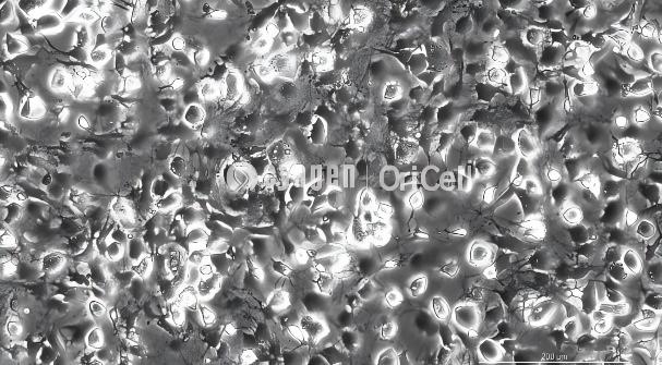
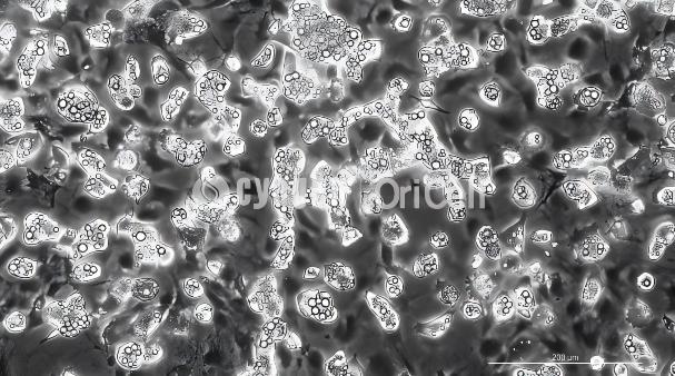
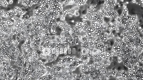
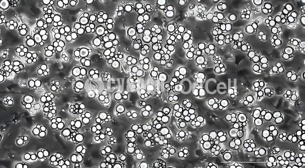
Images showing the induction process: From top to bottom: day 3, day 6, day 11, and day 15 of induction.
Staining and Identification
Oil Red O staining is used to detect red lipid droplets and verify adipogenic differentiation capacity.
Oil Red O Staining Procedure
- Termination of induction and washing: Aspirate the adipogenic induction medium, and gently wash the cells 2–3 times with pre-cooled 1× PBS to remove residual medium.
- Cell fixation: Add fixative to each well and fix at room temperature for 30 min.
- Staining reaction: Aspirate the fixative. After washing twice with 1× PBS, add Oil Red O working solution to cover the cells and stain at room temperature for 30 min.
- Termination of staining and washing: Aspirate the staining solution. Wash 2–3 times with 1× PBS until no floating background color remains, then add 1× PBS to keep the cells moist.
Staining result: The presence of round or oval red lipid droplets of varying sizes in the cytoplasm indicates adipogenic differentiation. A large number of densely distributed lipid droplets indicates successful differentiation. In contrast, no red lipid droplets or only scattered tiny particles indicates unsuccessful differentiation.
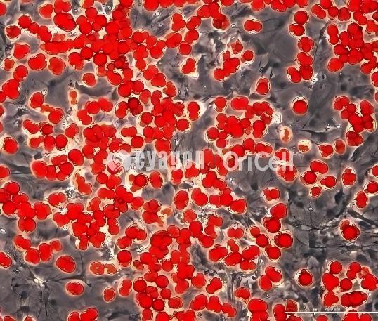
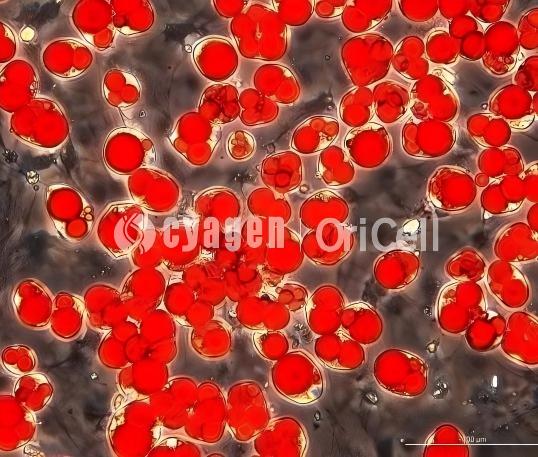
Oil Red O staining result, day 15.
Chondrogenic Differentiation
Core Steps for Chondrogenic Differentiation of Tendon Stem Cells
- • Cell counting: After digestion and centrifugation, discard the supernatant and add 1 mL of chondrogenic induction basal medium to resuspend the cells. Count the cell suspension and dilute it to a cell concentration of approximately 3 × 10⁵ cells/mL.
- • Cell seeding: After centrifugation, discard the supernatant and add 1 mL of chondrogenic induction basal medium to resuspend the cells. Centrifuge at 150 × g for 4 min. Discard the supernatant, add 1 mL of complete chondrogenic induction medium to resuspend the cells, and centrifuge again at 150 × g for 4 min. After centrifugation, do not shake or pipette the cell pellet. Carefully loosen the centrifuge tube cap to allow gas exchange, and place the tube in a 37°C, 5% CO₂ incubator.
- • Induction initiation: Replace with fresh chondrogenic differentiation induction medium every 2–3 days, using 0.5–1 mL per tube. After each medium change, gently tap the cell pellet to allow it to detach from the tube wall and remain suspended. Loosen the tube cap and continue induction at 37°C with 5% CO₂. During induction, the surface of the cell pellet will become smooth and gel-like. Continue induction until a cartilage pellet with a diameter of 1.5–2 mm forms in the tube, then proceed with paraffin sectioning. For preparation of the kit and detailed induction steps, please refer to the product manual.
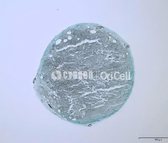
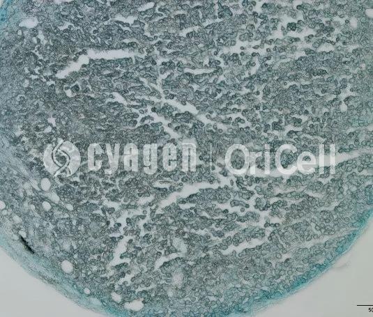
Images showing the induction process, day 21.
Staining and Identification
Paraffin sectioning followed by Alcian Blue staining is used for identification. The stained regions indicate acidic mucopolysaccharides within cartilage tissue.
Experimental FAQ
Q1: Why should cell density be controlled during osteogenic differentiation?
A: Excessively high confluence may cause cell detachment. We recommend controlling cell density at the early stage of induction.
Q2: How can osteogenic cell detachment and calcium nodule loss be reduced in the later stage of induction?
A: In the later stage of osteogenic induction, half-medium changes every 2 days can be used to reduce osteoblast detachment and calcium nodule loss.
Q3: Should the medium be prewarmed before medium changes?
A: Yes. The medium should be prewarmed before every medium change.
Q4: Why should calcium-containing buffers be avoided during Alizarin Red staining?
A: Calcium-containing buffers, such as PBS containing Ca²⁺, should be avoided to prevent nonspecific binding. After staining, samples should be washed thoroughly to reduce background interference.
Q5: What should be done if Alizarin Red staining is too intense?
A: Shorten the staining time or reduce the Alizarin Red concentration, for example to 0.1%.
Q6: How can calcium nodule detachment be avoided before Alizarin Red staining?
A: Avoid vigorous shaking of the culture plate before fixation.
Q7: What cell status is recommended for osteogenic differentiation and staining?
A: Low-passage cells in good condition should be used.
Q8: How should medium changes be performed during adipogenic induction?
A: Medium changes during induction should be gentle. We recommend adding induction medium slowly along the side wall of the culture vessel to prevent cell detachment or edge curling.
Q9: When should Solution A induction be stopped during adipogenic differentiation?
A: When a large number of small lipid droplets are observed, Solution A induction should be stopped and the cells should be switched to Solution B for maintenance. For example, in the image showing day 6 of induction, the culture can be switched to Solution B for subsequent maintenance until the lipid droplets appear pomegranate seed-like.
Q10: Why must medium changes be performed on time during adipogenic induction?
A: Medium changes should be performed promptly to prevent poor cell condition or cell death caused by insufficient nutrients.
Q11: How should Oil Red O working solution be prepared?
A: Mix Oil Red O stock solution with distilled water at a ratio of 3:2. After mixing, centrifuge at 250 × g for 4 min and use the supernatant. Alternatively, the mixture can be filtered before use.
Q12: How can high background after Oil Red O staining be reduced?
A: Wash thoroughly after Oil Red O staining or shorten the staining time.
Q13: Why do lipid droplets appear blurred?
A: Unclear lipid droplet boundaries may be caused by excessive differentiation, which can lead to lipid droplet fusion and rupture.
Q14: What should be considered when washing after Oil Red O staining?
A: During washing, remove impurities such as cell debris or insoluble staining solution particles, as these impurities may affect staining quality.
Q15: How should complete chondrogenic induction medium be prepared?
A: Complete chondrogenic induction medium must be freshly prepared before use.
Q16: Why should the cell pellet not be disturbed during the first 24 h after chondrogenic seeding?
A: The cell pellet should not be shaken during the first 24 h after seeding, as this period is critical for stable pellet formation.
Q17: Should the medium be prewarmed before chondrogenic medium changes?
A: Yes. The medium should be prewarmed before every medium change.
OriCell Featured Products
| Type | Product Name | Cat. No. | Size |
|---|---|---|---|
| Primary Cells | SD Rat Tendon Stem Cells | RASTA-01001 | 1 × 10⁶ |
| Culture Medium | Complete Medium For Rat Tendon Stem Cells | RAXTA-90011 | 100 mL |
| Induction Kit | Osteogenic Differentiation Medium For Rat Tendon Stem Cells | RASTA-90021 | 100 mL |
| Induction Kit | Adipogenic Differentiation Medium For Rat Tendon Stem Cells | RASTA-90031 | 100 mL |
| Induction Kit | Chondrogenic Differentiation Medium For Rat Tendon Stem Cells | RASTA-90041 | 100 mL |
About Cyagen OriCell
Cyagen OriCell is a Cyagen brand focused on the research and development of cell biology products, including stem cells, primary cells, and cell lines, as well as cell culture reagents and technical services. Serving universities, research institutes, hospitals, CROs, and CDMOs worldwide, Cyagen OriCell has accumulated extensive expertise in cell isolation and culture. The team has developed "spatial replication" culture technology to rapidly establish growth-supportive environments, and runs an Antibiotic-Free process grounded in strict environmental, materials, and personnel controls. Cyagen OriCell provides end-to-end solutions—from MSC isolation and identification to directed differentiation and assay services.
Cyagen OriCell's offerings are cited in over 10,000 publications, with a cumulative impact factor exceeding 90,000 and more than 160,000 citations, and the team has supported more than 3,000 research groups. Products are used by tens of thousands of customers across dozens of countries and regions.

