
 EN
EN
Why Are Mineralized Nodules Absent During Osteogenic Differentiation of MC3T3-E1 Cells?
At Cyagen, we often receive questions from researchers performing osteogenic differentiation experiments with MC3T3-E1 cells: “Why did my cells fail to form mineralized nodules after induction?” This is also a question frequently discussed on Reddit and other lab forums, where researchers compare induction conditions, staining results, and troubleshooting experiences for MC3T3-E1 osteogenic differentiation.
The MC3T3-E1 cell line is derived from the calvaria, or skull bone, of C57BL/6 mice and has the ability to differentiate into osteoblast-like cells. Under osteogenic induction conditions, cells typically undergo three progressive stages: proliferation, extracellular matrix maturation, and mineralization.
This differentiation process is commonly induced using a combination of key supplements:
- • Ascorbic acid, which promotes collagen synthesis and extracellular matrix maturation;
- • β-glycerophosphate, which provides phosphate ions required for mineral deposition;
- • Dexamethasone, which stimulates the expression of osteogenic markers.
During the differentiation process, researchers usually evaluate osteogenic progression through multiple approaches:
- • Alkaline phosphatase (ALP) staining or quantitative analysis to assess early-stage osteogenic differentiation;
- • Alizarin Red staining to detect calcium deposition and mineralized nodule formation at the late stage;
- • qRT-PCR analysis of osteogenic marker genes, including Runx2, osteocalcin (OCN), and bone sialoprotein (BSP), for comprehensive evaluation.
However, in some experiments, MC3T3-E1 cells may show limited or no mineralized nodule formation after induction. This can be caused by several factors, including induction conditions, cell status, culture environment, and experimental handling.
In this article, Cyagen scientists will share common causes behind unsuccessful mineralization and provide practical troubleshooting suggestions to help researchers optimize their MC3T3-E1 osteogenic differentiation experiments.
Learn More About MC3T3-E1 Osteogenic Differentiation
To support researchers performing osteogenic differentiation studies, Cyagen scientists have prepared a practical experimental guide covering key steps, optimization tips, and commonly encountered issues during MC3T3-E1 induction.
Interested in detailed protocol guidance or experimental resources? Contact our scientific team to request more information.
01 MC3T3-E1 Cell Line and Subclone Selection
The MC3T3-E1 Family
The MC3T3-E1 family consists of multiple subclones with distinct characteristics. These subclones all originate from the same parental cell source, but during long-term passaging and selection, they developed different functional properties and differentiation capacities.
| Characteristic | MC3T3-E1 Subclone 4 | MC3T3-E1 Subclone 14 | MC3T3-E1 Subclone 24 | MC3T3-E1 Subclone 30 |
|---|---|---|---|---|
| Differentiation potential | High | High | Low | Low |
| Mineralization capacity | Forms a well-mineralized extracellular matrix | Forms ECM, but the mineralization pattern may be more diffuse | Does not form mineralized ECM | Does not form mineralized ECM |
| Osteogenic markers, such as BSP and OCN | Highly expressed | Highly expressed | Low expression | Low expression |
| PTH1R functional response | Responsive; can produce cAMP | Poor or no response | Poor or no response | Information unavailable |
Different MC3T3-E1 subclones show marked differences in osteogenic differentiation capacity. Selecting the correct subclone is therefore critical for experimental success.
Recommended subclones: MC3T3-E1 Subclone 14 or Subclone 4. In the presence of ascorbic acid and inorganic phosphate, these subclones show a high level of osteogenic differentiation capacity and can form a well-mineralized extracellular matrix in approximately 10–14 days.
02 MC3T3-E1 Osteogenic Differentiation Experimental Guide
Experimental Materials
- Cell line: OriCell MC3T3-E1 Subclone 14 Mouse Calvarial Preosteoblast Subclone 14 Cell Line (Cat. No. M7-0201).
Tip: Low-passage cells are recommended because they usually show better differentiation potential. - Growth medium: OriCell Complete Medium For MC3T3-E1 Subclone 14 Cell Line (Cat. No. CMM7-0201), containing basal medium and Uruguayan fetal bovine serum.
- Osteogenic induction medium: OriCell Osteogenic Differentiation Medium For Mouse MC3T3-E1 Cells (Cat. No. MUXMT-90021), containing basal medium, New Zealand fetal bovine serum, osteogenic induction supplements, Alizarin Red staining solution, and gelatin.
Tip: Fetal bovine serum should be screened to select a batch suitable for osteogenic induction. - Other reagents: PBS, 0.25% trypsin-EDTA, 4% paraformaldehyde, and BCIP/NBT ALP color development kit.
Experimental Procedure
Stage 1: Cell Culture and Seeding
- Culture vessel coating, optional but strongly recommended:
Because the induction period is long, usually 2–4 weeks, cells may curl at the edges and detach. We recommend covering the bottom of the culture vessel with 0.1% gelatin, incubating it in the incubator for 30 min, aspirating the gelatin solution, and allowing the vessel to air-dry before use. - Cell seeding:
Use MC3T3-E1 Subclone 14 cells in the logarithmic growth phase and in good condition. After trypsin digestion, count the cells. - Seeding density:
We recommend seeding at a density of 2–3 × 10⁴ cells/cm², for example in 12-well or 24-well plates. High-density seeding helps cells reach confluence more quickly and enter the differentiation stage. - Cell culture:
Culture the cells in MC3T3-E1 Subclone 14 growth medium at 37°C in a 5% CO₂ incubator.
Stage 2: Osteogenic Induction
We recommend setting up both a control group, cultured in regular growth medium, and an experimental group, cultured in osteogenic induction medium.
- Observe cell confluence. When cells reach 80%–100% confluence, induction can be started.
- Aspirate the MC3T3-E1 Subclone 14 growth medium.
- Experimental group: Add prewarmed osteogenic differentiation complete medium, using OriCell Osteogenic Differentiation Medium For Mouse MC3T3-E1 Cells.
- Control group: Add cell growth medium, using OriCell Complete Medium For MC3T3-E1 Subclone 14 Cell Line.
- Replace with fresh medium every 48–72 h. Medium changes should be performed gently. Add medium slowly along the wall of the well to avoid dislodging the cell monolayer.
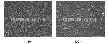
MC3T3-E1 cell line | Reference confluence before induction
Stage 3: Staining and Characterization
Detection should be performed at different time points according to the differentiation process.
1. Alkaline Phosphatase (ALP) Detection
Early-stage marker, usually detected on days 7–14.
ALP staining, qualitative detection:
- Aspirate the medium, wash with PBS, and fix with 4% paraformaldehyde for 30 min.
- After washing with PBS, add BCIP/NBT staining working solution and incubate at room temperature protected from light for 30 min. Positive cells appear blue-purple.
2. Mineralized Nodule Detection
Late-stage marker, usually detected on days 14–21.
Alizarin Red staining, qualitative detection:
- Fixation: Same as the ALP staining procedure.
- Staining: Add 1% Alizarin Red staining solution and stain at room temperature for 10–15 min.
- Washing: Wash repeatedly with PBS or distilled water to remove background staining.
- Observation: Observe under a microscope. Orange-red or dark red nodules indicate mineralized nodules.
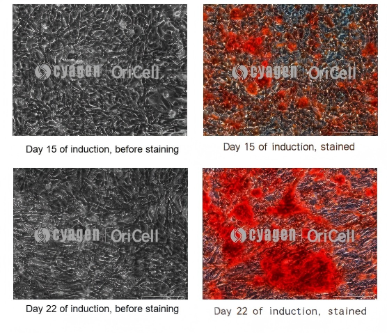
MC3T3-E1 cell line | Alizarin Red staining at the late stage of osteogenic induction
Expected Results
| Evaluation Item | Expected Result |
|---|---|
| Morphological changes | Approximately 1 week after induction, cells gradually change from an elongated spindle-like morphology to a cobblestone-like morphology. After 2 weeks, opaque white nodules, representing early mineralized nodules, may be observed on the cell layer. |
| ALP staining | On days 7–14, the induction group shows a large number of blue-purple positive cells, while the control group is generally negative. |
| Alizarin Red staining | On days 14–21, orange-red mineralized nodules of different sizes can be observed in the induction group, while no red staining is observed in the control group. |
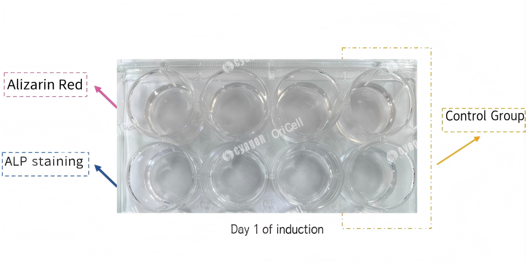
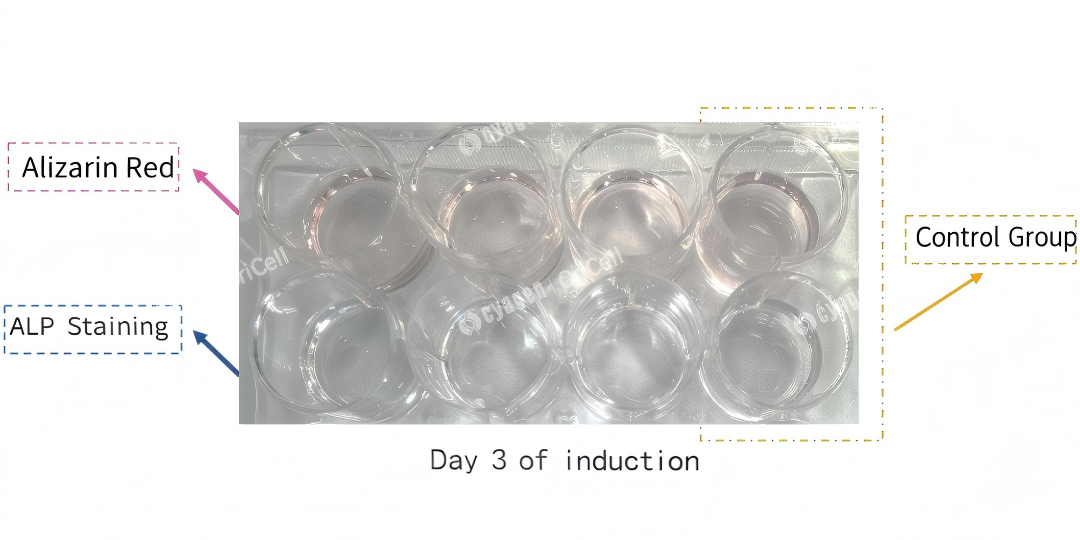
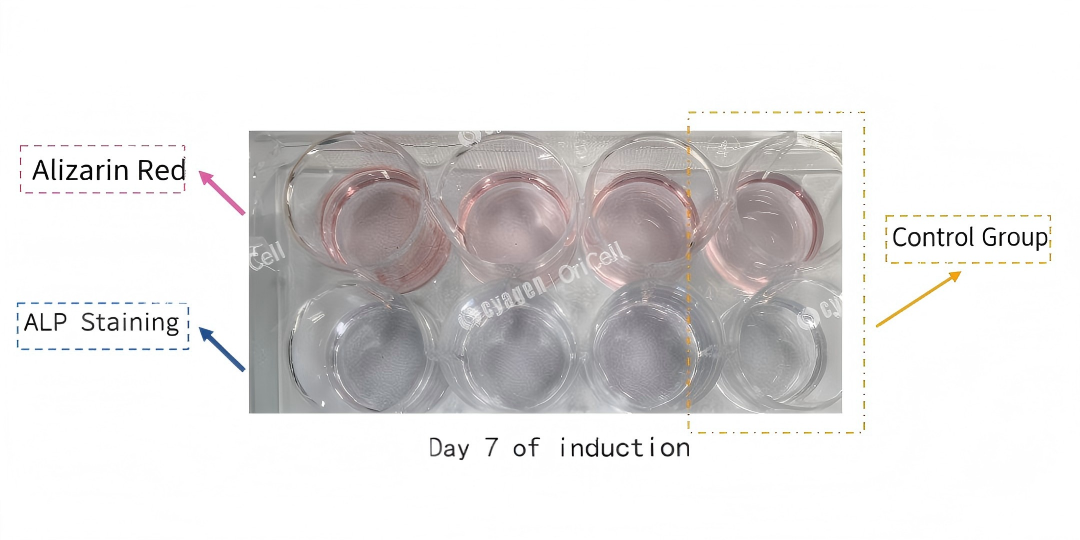
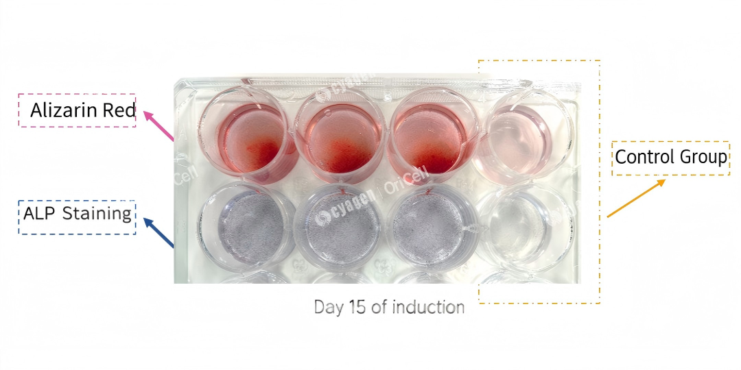
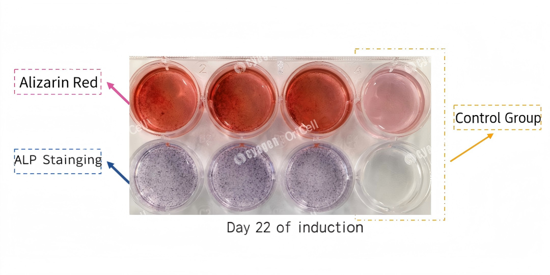
Full-cycle induction staining in a plate. The first three wells are the induction group, and the last well is the negative control group. The first row shows Alizarin Red staining, and the second row shows ALP staining.
Experimental FAQ: Key Considerations and Troubleshooting
Q1: Why is low-passage MC3T3-E1 important for osteogenic differentiation?
A: Low-passage cells should be used whenever possible. High-passage cells may lose differentiation potential. Before starting the experiment, make sure the cells are in good condition and free from contamination.
Q2: Why does uneven cell seeding affect differentiation results?
A: The cell suspension must be mixed thoroughly before seeding. Uneven seeding can lead to inconsistent differentiation and a patchy staining pattern.
Q3: How can cell detachment be reduced during long-term induction?
A: Gelatin coating is strongly recommended for long induction periods. Coating the culture vessel can help reduce edge curling and cell detachment.
Q4: How should medium changes be performed to avoid disrupting the cell layer?
A: Add medium slowly along the wall of the well during medium changes, or leave a small amount of old medium as a buffer. Avoid directly flushing the cell monolayer.
Q5: How should the osteogenic induction medium be prepared and handled?
A: When using OriCell Osteogenic Differentiation Medium For Mouse MC3T3-E1 Cells, freshly preparing the medium before use is recommended to maintain the stability and effectiveness of the osteogenic induction supplements. The medium should be prewarmed before use.
Q6: What should be done if Alizarin Red or ALP staining shows dirty background or precipitates?
A: Alizarin Red staining solution can be centrifuged before use to remove precipitates. After staining, the samples should be washed thoroughly.
Q7: Why is staining weak or why are mineralized nodules absent?
A: Possible causes include insufficient induction time, as standard induction usually requires 2–3 weeks; loss of differentiation potential due to high passage number; or failure of induction reagents due to expiration or improper storage.
Q8: What can be done if staining is weak or no mineralized nodules are observed?
A: Extend the induction period as appropriate, check reagent expiration dates, and set up a positive control reagent or positive control condition.
Q9: Why does uneven staining occur?
A: Uneven staining is usually caused by uneven cell seeding or local cell detachment during the induction process.
03 Cyagen 3T3 Cell Product and Service Recommendations
Cyagen provides a one-stop solution for 3T3 cell research, including in-stock cell lines that have passed STR authentication and mycoplasma testing, cell culture reagents such as growth medium, differentiation induction medium, cryopreservation medium, and dissociation solution, as well as technical services for cell gene modification, including gene knockout, point mutation, gene knock-in, overexpression, and knockdown. Routine cell function and characteristic testing services are also available.
OriCell Related Product Recommendations
KO Cell Model Recommendations (Selected Examples)
| Cell Name | Gene Name | NCBI ID | Service No. |
|---|---|---|---|
| 3T3-L1 | Ctbp1 | 13016 | SY-KO-00107 |
| MC3T3 | Piezo1 | 234839 | SY-KO-00330 |
| MC3T3-E1 | Ccdc134 | 76457 | SY-KO-00470 |
| MC3T3-E1 | Fam20b | 215015 | SY-KO-00408 |
| MC3T3-E1 | Lgr5 | 14160 | SY-KO-00342 |
| NIH/3T3 | Fbxl12 | 30843 | SY-KO-00410 |
| NIH/3T3 | Rpgrip1l | 244585 | SY-KO-00422 |
| NIH/3T3 | Smo | 319757 | SY-KO-00290 |
| NIH3T3 | 4933427D06Rik | 232217 | SY-KO-00084 |
About Cyagen OriCell
Cyagen OriCell is a Cyagen brand focused on the research and development of cell biology products, including stem cells, primary cells, and cell lines, as well as cell culture reagents and technical services. Serving universities, research institutes, hospitals, CROs, and CDMOs worldwide, Cyagen OriCell has accumulated extensive expertise in cell isolation and culture. The team has developed "spatial replication" culture technology to rapidly establish growth-supportive environments, and runs an Antibiotic-Free process grounded in strict environmental, materials, and personnel controls. Cyagen OriCell provides end-to-end solutions—from MSC isolation and identification to directed differentiation and assay services.
Cyagen OriCell's offerings are cited in over 10,000 publications, with a cumulative impact factor exceeding 90,000 and more than 160,000 citations, and the team has supported more than 3,000 research groups. Products are used by tens of thousands of customers across dozens of countries and regions.

