
 EN
EN
RAW264.7 Cells Differentiating Unexpectedly? A Troubleshooting Guide for Maintaining Cell Characteristics
RAW264.7 cells are a mouse-derived monocyte/macrophage cell line widely used in immunology, inflammatory response studies, tumor microenvironment research, and drug screening applications. This is also a recurring topic in Reddit discussions and lab forums, where researchers often ask why RAW264.7 cells suddenly change morphology or show stronger adhesion during routine culture. Due to their easy maintenance and high transfection efficiency, RAW264.7 cells have become one of the most widely used macrophage models in research laboratories.
However, maintaining RAW264.7 cells with consistent characteristics can sometimes be challenging. Under certain culture conditions, RAW264.7 cells may undergo unwanted differentiation, accompanied by changes in morphology, adhesion properties, and cellular responses. These changes may affect experimental reproducibility and the reliability of downstream assays, especially in studies involving immune activation, inflammation, or macrophage-related mechanisms.
Why do RAW264.7 cells differentiate unexpectedly? What factors contribute to changes in cell state, and how can researchers maintain stable RAW264.7 cell characteristics during culture?
In this guide, OriCell scientists summarize key factors that may influence RAW264.7 cell differentiation and share practical troubleshooting strategies to help researchers optimize cell culture conditions and maintain reliable experimental performance.
Cell Overview
| Parameter | Information |
|---|---|
| Product name | RAW 264.7 Mouse Monocyte Macrophage Leukemia Cell Line |
| Abbreviation | RAW264.7 |
| Alternative names | RAW264, RAW2647, RAW264.7, RAW-264.7, Raw 264.7, Raw264.7 |
| Culture system | DMEM + 10% FBS, 37°C, 5% CO₂ |
| Cell morphology | Macrophage-like morphology; adherent growth |
| Doubling time | 24–48 h |
| Recommended split ratio | 1:3 to 1:6 |
| OriCell cell catalog number | M3-0101 |
| OriCell Complete Medium for Cell Line | CMM3-0101, 500 mL |
The RAW264.7 mouse monocyte/macrophage leukemia cell line was isolated from tumor tissue of a male BALB/c mouse induced by Abelson murine leukemia virus. This cell line is easy to culture and passage, sensitive to RNA interference, and highly efficient for DNA transfection. It is commonly used as a transfection host cell and for replication studies of murine norovirus. RAW264.7 cells are negative for surface sIg, Ia, and Thy-1.2 antigens.
RAW264.7 cells can be used in studies of oxidative stress, inflammation, antibacterial activity, and related biological processes. In immunology research, they are widely used to investigate macrophage immune response mechanisms, such as cytokine production and function. In inflammation-related studies, they can help model inflammatory responses in vitro and support anti-inflammatory drug screening.
1. What Is the Typical Morphology of RAW264.7 Cells?
Under routine culture conditions and in the absence of induced differentiation, RAW264.7 cells mainly display two morphologies: loosely adherent spindle-shaped cells and round or cuboidal cells. As cell density increases in the culture system, some adherent cells may undergo morphological changes, including cell body contraction, rounding, or cell aggregate formation. Some cells may detach from the culture surface and enter a suspension state.
Tip: These suspended cells can still retain biological activity and should be kept during passaging. They can be collected by centrifugation at 250 × g for 4 min. After resuspending the resulting cell pellet, the cells can continue to be used for subsequent culture.
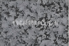
100X
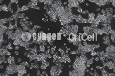
200X
Good morphology of RAW264.7 cells under the microscope
2. What Does RAW264.7 Cell Morphology Look Like After Differentiation?
After differentiation or polarization, RAW264.7 cells may shift from loosely adherent spindle-shaped, round, or cuboidal morphology to large, flattened, irregular polygonal cells or cells with multiple protrusion-like structures. Their adhesion capacity becomes significantly stronger.
- •Clear filamentous pseudopodia may be observed at the cell edges.
- •The cytoplasmic spreading area increases, and the cells become flattened.
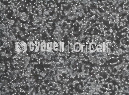
40X
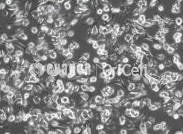
100X
Polarized morphology of RAW264.7 cells under the microscope
3. How Can RAW264.7 Cells Be Handled to Reduce Differentiation?
Gentle Pipetting-Based Passaging Method
- During passaging, use a pipette or serological pipette to aspirate part of the culture supernatant, leaving a small amount of supernatant in the flask or dish to facilitate gentle pipetting. For example, leave only 2–3 mL of supernatant in a 6 cm culture dish.
- If a large number of suspended cells are observed under the microscope, collect all the supernatant and centrifuge it together with the cells detached by pipetting to avoid cell loss.
- Use a 1 mL disposable pipette tip or serological pipette to aspirate the culture supernatant and pipette directly across the bottom of the culture vessel several times.
- Avoid vigorous pipetting and excessive bubble formation, as this may damage the cells. Differentiated cells that cannot be detached by pipetting should be discarded.
- Transfer the cell suspension into a 15 mL centrifuge tube.
- Wash the culture vessel once with PBS, using approximately 3 mL for a 6 cm dish and approximately 6 mL for a 10 cm dish, to collect residual cells.
- Centrifuge all collected cell suspensions at 250 × g for 4 min.
- After centrifugation, remove the supernatant.
- Add 2 mL of complete medium to the 15 mL centrifuge tube, and gently pipette the cell pellet to fully resuspend and mix the cells.
- Seed the cells into an appropriate culture vessel at a density of (4–6) × 10⁴ viable cells/cm².
- Gently mix the cells evenly and place them in a CO₂ incubator at 37°C, 5% CO₂, and saturated humidity.
- On the day after passaging, observe the cell condition. If many floating cells are present and appear to be in poor condition, change the medium.
- Replace with fresh medium every 3 days. When cell confluence reaches above 85%, the cells should be passaged or cryopreserved.
Experimental FAQ
Q1: Are suspended RAW264.7 cells still usable during routine culture?
A: Yes. Some RAW264.7 cells may detach and enter suspension as cell density increases. These suspended cells can still retain biological activity and should be kept during passaging rather than discarded.
Q2: How should suspended RAW264.7 cells be collected?
A: Suspended cells can be collected by centrifugation at 250 × g for 4 min. After centrifugation, resuspend the cell pellet and continue culture.
Q3: What morphology suggests that RAW264.7 cells may have differentiated or polarized?
A: RAW264.7 cells may become large, flattened, and irregularly polygonal, or develop multiple protrusion-like structures. They may also show stronger adhesion, clear filamentous pseudopodia at the cell edges, and increased cytoplasmic spreading.
Q4: Why should vigorous pipetting be avoided during passaging?
A: Vigorous pipetting can generate excessive bubbles and mechanical stress, which may damage RAW264.7 cells and reduce cell viability.
Q5: What should be done with differentiated RAW264.7 cells that cannot be detached by pipetting?
A: Differentiated cells that remain strongly attached and cannot be detached by gentle pipetting should be discarded.
Q6: Why should all supernatant be collected if many suspended cells are present?
A: If many suspended cells are present, discarding the supernatant may result in substantial cell loss. The supernatant should be collected and centrifuged together with the pipetted cell suspension.
Q7: Why is a culture dish recommended for RAW264.7 cell culture?
A: Compared with culture flasks, culture dishes are easier to pipette due to differences in physical space and surface tension. Their open structure allows pipette tips to operate at a lower angle, reducing fluid shear stress and minimizing damage to weakly adherent cells.
Q8: When should RAW264.7 cells be passaged or cryopreserved?
A: When cell confluence reaches above 85%, RAW264.7 cells should be passaged or cryopreserved.
OriCell Featured Products
| Type | Product Name | Cat. No. | Size |
|---|---|---|---|
| Cell Line | RAW 264.7 Mouse Monocyte Macrophage Leukemia Cell Line | M3-0101 | 1 × 10⁶ |
| Cell Culture Media | Complete Medium For RAW 264.7 Cell Line | CMM3-0101 | 500 mL |
About Cyagen OriCell
Cyagen OriCell is a Cyagen brand focused on the research and development of cell biology products, including stem cells, primary cells, and cell lines, as well as cell culture reagents and technical services. Serving universities, research institutes, hospitals, CROs, and CDMOs worldwide, Cyagen OriCell has accumulated extensive expertise in cell isolation and culture. The team has developed "spatial replication" culture technology to rapidly establish growth-supportive environments, and runs an Antibiotic-Free process grounded in strict environmental, materials, and personnel controls. Cyagen OriCell provides end-to-end solutions—from MSC isolation and identification to directed differentiation and assay services.
Cyagen OriCell's offerings are cited in over 10,000 publications, with a cumulative impact factor exceeding 90,000 and more than 160,000 citations, and the team has supported more than 3,000 research groups. Products are used by tens of thousands of customers across dozens of countries and regions.

