
 EN
EN
Primary Chondrocyte Isolation Protocol: Step-by-Step Enzymatic Digestion and Culture Method
Cartilage tissue is composed of chondrocytes embedded within an extracellular matrix rich in fibers and structural components. Although cartilage has relatively limited regenerative capacity compared to other tissues, it contains a population of proliferative immature cells that can differentiate into chondroblasts and eventually mature chondrocytes, forming a stable cartilage matrix. Mature chondrocytes reside within lacunae and are responsible for maintaining cartilage homeostasis.
In histological structure, immature chondrocytes are typically located in the superficial cartilage zone, appearing as small, oval-shaped cells aligned parallel to the cartilage surface. As cells progress into deeper layers, they become larger and more spherical, with round or oval nuclei, lightly stained cytoplasm, and varying amounts of lipid droplets.
Primary chondrocytes derived from neonatal animals are widely used due to their high viability, superior yield, and robust biological activity. Cyagen OriCell chondrocytes are isolated from cartilage tissue using a combined collagenase and neutral protease enzymatic digestion method, ensuring high purity and functionality for downstream applications.
In vitro cultured chondrocytes are essential for studying cartilage physiology, drug response, and pathological mechanisms under disease conditions such as osteoarthritis. This article provides a practical, step-by-step protocol for primary chondrocyte isolation and extraction, designed to support both basic research and translational studies.
Primary Chondrocyte Isolation and Extraction Procedure
Experimental Materials
Sample
Neonatal mice within 7 days after birth.
Reagents
OriCell Complete Medium For Rat Costal Chondrocytes, OriCell 0.1% Collagenase Type I, OriCell 0.25% Neutral Protease, and OriCell Phosphate-Buffered Saline Solution (1X), 100 mL.
Consumables and instruments
10 cm culture dishes, 6 cm culture dishes, ophthalmic forceps (4), ophthalmic scissors (2), disposable pipettes, and sterile dissection trays.
Workflow Overview
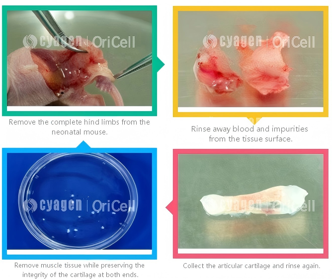
Experimental Procedure
- Euthanize the mouse by CO₂ inhalation, then immerse it in 75% ethanol for 2–3 min.
- In a clean bench, remove the complete hind limbs of the mouse and place them into a culture dish containing PBS with 2% penicillin-streptomycin. Wash to remove surface blood and impurities.
- Using clean ophthalmic forceps and ophthalmic scissors, remove the muscle tissue from the femur and tibia while preserving the integrity of the cartilage at both ends.
- Separate the articular cartilage from the bone, and thoroughly remove any attached muscle and fascia from the cartilage.
- Collect the cartilage in a 6 cm culture dish and mince the tissue with ophthalmic scissors into pieces of approximately 0.5 mm³.
- Add a mixed solution of Collagenase Type I and Neutral Protease. Place the culture dish in a 37°C incubator and digest for 6–8 h. Every 1–2 h, remove the dish and gently pipette the tissue using a disposable pipette to promote digestion.
- After digestion is complete, terminate the digestion using chondrocyte complete medium, then centrifuge.
- Centrifuge at 300 × g for 5 min. Discard the supernatant, resuspend the pellet in PBS, and centrifuge again.
- Resuspend the cell pellet in chondrocyte-specific complete medium and seed the cells. Place the seeded culture dish in a humidified incubator at 37°C with 5% CO₂.
- Observe the cells the next day to determine whether there are many cell fragments and dead cells. A half-medium change or complete medium change may be considered as appropriate.
- After 48 h, cell confluence can reach 90%. At this stage, the cells are three-dimensional and translucent, with spindle-shaped or polygonal morphology, and can be passaged at a ratio of 1:3.
Experimental FAQ
Q: Why should cartilage tissue be minced into approximately 0.5 mm³ pieces?
A: Tissue fragments of this size help support efficient enzymatic digestion while minimizing cell damage. If the pieces are too large, enzymatic digestion may be incomplete; if they are too small, the cells may be damaged.
Q: Why should the tissue be pipetted every 1–2 h during enzymatic digestion?
A: Gentle pipetting during digestion helps promote tissue dissociation and improves digestion efficiency.
Q: Why is the cell pellet washed and centrifuged again after digestion?
A: This step helps remove residual enzymes, which may otherwise affect subsequent cell attachment.
Q: How should culture conditions be optimized for primary chondrocytes?
A: Using a medium with a high serum concentration, such as 10% fetal bovine serum plus nutritional factors, can help improve cell attachment and proliferation.
Q: What does OriCell chondrocyte complete medium contain?
A: OriCell chondrocyte complete medium contains a basal medium suitable for the growth of primary chondrocytes, OriCell premium fetal bovine serum, and other supplements required for cell growth. It can support the long-term maintenance of primary chondrocytes in good in vitro growth condition.
Cell Characterization: OriCell Chondrocyte Quality Control Criteria
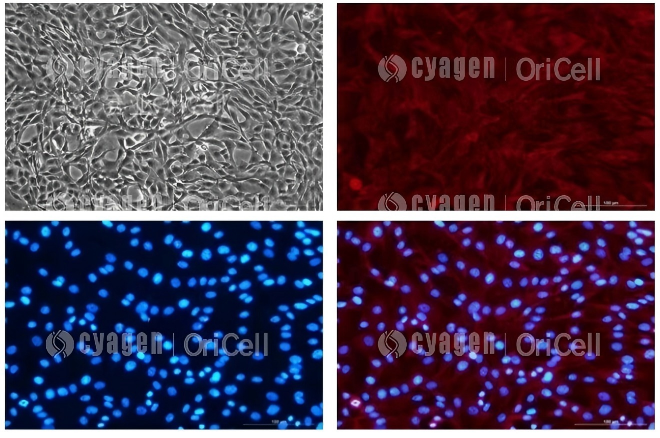
- Cell identification: Collagen II positivity >90% by immunofluorescence detection.
- Cells pass bacterial, fungal, mycoplasma, and endotoxin testing.
- Cells pass post-thaw viability testing, with a post-thaw survival rate >90%.
From left to right and top to bottom, the images show bright-field, fluorescence, nuclear staining, and merged images of mouse chondrocytes.
Cyagen OriCell In-Stock Chondrocyte Product Recommendations
Supported by a mature and reliable primary cell isolation platform, Cyagen OriCell can provide high-quality in-stock primary chondrocytes and supporting culture media. In addition, we can customize fluorescently labeled primary cells according to customer needs and isolate primary chondrocytes from other species, such as mouse, to fully support your research requirements.
| Type | Product Name | Cat. No. | Size |
|---|---|---|---|
| Primary Cells | OriCell SD Rat Knee Articular Chondrocytes | RASKJ-02071 | 1*10^6/vial |
| Primary Cells | OriCell SD Rat Thoracic Chondrocytes | RASST-02071 | 1*10^6/vial |
| Primary Cells | OriCell SD Rat Costal Chondrocytes | RASRB-02071 | 1*10^6/vial |
| Cell Culture Media | OriCell Complete Medium For Rat Costal Chondrocytes | RASRB-90011 | 500mL |
| Cell Culture Media | OriCell Complete Medium For Rat Thoracic Chondrocytes | RASST-90011 | 500mL |
| Reagents | OriCell Phosphate-Buffered Saline Solution (1X) | PBS-10001 | 500mL |

