
Umbilical Cord Mesenchymal Stem Cells (UCMSCs): Benefits, Isolation and Culture Guide
Umbilical cord mesenchymal stem cells (UCMSCs) are one of the most widely used mesenchymal stem cell sources in regenerative medicine due to their accessibility, robust proliferative capacity, and low immunogenicity. Understanding their biological characteristics is essential for selecting the right cell source, while mastering appropriate isolation and culture techniques is critical for obtaining high-quality primary cells.
In this guide, we first introduce the origin and biological advantages of UCMSCs before reviewing commonly used isolation and primary culture methods, providing researchers with both the scientific background and practical laboratory guidance needed for UCMSC research.
Tissue Origin of UCMSCs
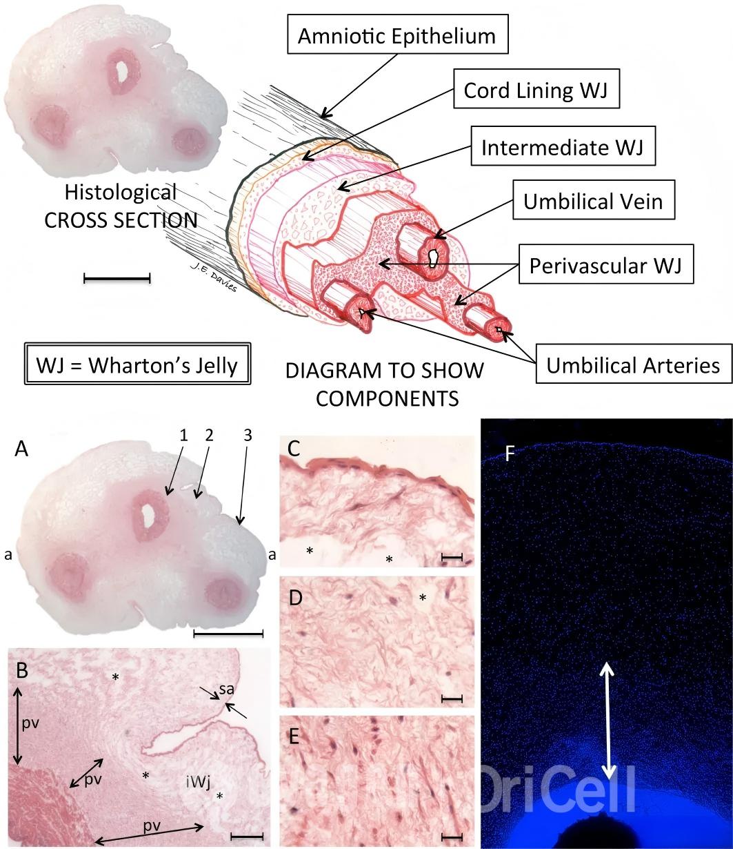
Umbilical cord mesenchymal stem cells are derived from a gelatinous connective tissue in the umbilical cord known as Wharton’s jelly. This tissue was first described in 1656 by the British physician and anatomist Thomas Wharton (1614–1673), after whom it was named. Anatomically, the umbilical cord consists of two umbilical arteries, one umbilical vein, and Wharton’s jelly, which surrounds all three umbilical vessels throughout the cord. One of the key functions of Wharton’s jelly is to protect the umbilical vessels from mechanical compression or twisting caused by external forces. Wharton’s jelly appears milky white and semi-transparent, and its major components include hyaluronic acid and chondroitin sulfate.
Because this tissue contains abundant mesenchymal stem cells, these cells are named according to their tissue of origin: umbilical cord mesenchymal stem cells (UCMSCs).
Three-dimensional structure of the umbilical cord (Stem Cells Translational Medicine 2017;6:1620–1630)
Advantages of UCMSCs
In the past, the umbilical cord was usually discarded as medical waste after birth. However, because umbilical cord tissue is rich in mesenchymal stem cells, it has become a valuable biological resource. Umbilical cord tissue is easy to obtain, free of major ethical concerns, and shares the key advantages of mesenchymal stem cells, including self-renewal, multilineage differentiation, homing capacity, and tissue repair potential. Compared with mesenchymal stem cells derived from adult tissues, UCMSCs are more primitive, exhibit stronger proliferative capacity, have lower immunogenicity, and show potent immunomodulatory activity. These advantages have made UCMSCs a highly valued cell source among stem cell researchers.
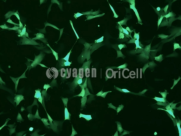
Microscopic image of UCMSC/GFP
It has been reported that in 2022, a total of 17 stem cell-based new drugs in China received implied clinical trial approval for IND applications from the Center for Drug Evaluation (CDE) of the National Medical Products Administration. These products were mainly intended for therapeutic research in hepatitis B virus-related liver cirrhosis, coronary heart disease, ankylosing spondylitis, burns, lupus nephritis, arthritis, and other indications. The main cell sources included MSCs derived from human bone marrow, adipose tissue, and umbilical cord tissue. Among them, research related to human umbilical cord mesenchymal stem cells accounted for nearly half of the total, underscoring their considerable potential.
UCMSC Isolation and Culture
Step 0: Material Preparation
- Sample: one umbilical cord collected within 8 hours, with a length of ≥5 cm. Storage temperature: 4°C.
- Reagents: 50 mL OriCell Complete Medium For Human Umbilical Cord Mesenchymal Stem Cells and 100 mL OriCell Phosphate-Buffered Saline Solution (1X).
- Consumables: one 50 mL sterile collection bottle, 5–10 50 mL centrifuge tubes, four 10 mL serological pipettes, and several T75 culture flasks.
Step 1: Umbilical Cord Processing
- Open the umbilical cord collection bottle aseptically and discard the preservation solution.
- Wash the umbilical cord in the collection bottle twice with OriCell Phosphate-Buffered Saline Solution (1X).
- Remove the umbilical cord with forceps and wash away residual peripheral blood from the cord surface using OriCell Phosphate-Buffered Saline Solution (1X).
- Cut off both ends tied with umbilical cord clamps or ties. For the middle segment of the cord, use toothed forceps and hemostatic forceps to remove residual blood from the umbilical vein and arteries. Wash the cord once again, and then cut it into small segments approximately 3 cm in length.
- Remove the one umbilical vein and two umbilical arteries, and isolate Wharton’s jelly.
- Use curved scissors to mince the Wharton’s jelly in the centrifuge tube into pieces of approximately 2–3 mm².
- Transfer the Wharton’s jelly into a 50 mL centrifuge tube and weigh 1 g of tissue pieces using an electronic balance.
- Add 10 mL of OriCell Complete Medium For Human Umbilical Cord Mesenchymal Stem Cells to the tissue pieces and mix well.
- Transfer the mixture into a T75 culture flask and distribute the tissue pieces evenly across the flask.
- Place the culture flask in a CO₂ incubator at 37°C, 5% CO₂, and saturated humidity.
Medium Change and Passaging Culture
Step 2: Medium Change
- Perform the first medium change on day 5 after completion of primary umbilical cord processing.
- Recover the culture medium and unattached tissue pieces from the culture flask as carefully as possible without disturbing the attached tissue pieces.
- Centrifuge the recovered tissue pieces and spent medium at 250 × g for 6 min, then discard the supernatant.
- Add 10 mL of fresh OriCell Complete Medium For Human Umbilical Cord Mesenchymal Stem Cells and reseed the material into a T75 culture flask.
- On day 7 after completion of the first medium change, perform the second half-medium change. This timing can be adjusted according to cell growth.
- Using a 10 mL serological pipette, recover half of the culture medium from the culture flask as carefully as possible without disturbing the attached tissue pieces.
- Add 5 mL of fresh OriCell Complete Medium For Human Umbilical Cord Mesenchymal Stem Cells to the T75 culture flask.
- After the second medium change, perform a half-medium change every 3 days until the cells are ready for passaging.
Step 3: P0-to-P1 Passaging
- After confirming that the cells are ready for passaging, remove floating tissue pieces and large tissue fragments. Pool and recover the tissue pieces and culture medium from the flask.
- Centrifuge at 600 × g for 5 min and collect the supernatant.
- Rinse the residual medium from the cells in the culture flask using OriCell Phosphate-Buffered Saline Solution (1X). Add 3 mL of OriCell 0.25% Trypsin-0.04% EDTA Solution to the T75 culture flask to digest the adherent cells.
- After digestion is complete, add 5 mL of the previously collected supernatant to the T75 flask to terminate digestion. Keep the remaining tissue fragment mixture in the centrifuge tube, label it, and store it in a 4°C refrigerator as a backup until cell cryopreservation is completed.
- Recover all cell suspensions, add OriCell Phosphate-Buffered Saline Solution (1X) to a final volume of 45 mL, and filter the suspension through a 100 μm cell strainer. Collect the filtrate, centrifuge at 250 × g for 6 min, and discard the supernatant.
- Resuspend the cell pellet in 30 mL of OriCell Phosphate-Buffered Saline Solution (1X). Take approximately 300 μL of the cell suspension for cell counting and viability assessment.
- According to the required seeding density, transfer the required volume of cell suspension, centrifuge at 250 × g for 6 min, and discard the supernatant. Add 25 mL of OriCell Complete Medium For Human Umbilical Cord Mesenchymal Stem Cells and seed the cells into one T175 culture flask.
- Place the culture flask in an incubator and culture at 37°C with 5% CO₂.
Step 4: Passaging After P1
- Prewarm the complete medium, PBS, and trypsin solution to 37°C.
- Aspirate the medium from the culture vessel. Wash the cells twice with PBS, using approximately 3 mL for a T25 flask and approximately 6 mL for a T75 flask. Perform this step gently and ensure complete washing, then aspirate the PBS.
- Add trypsin solution, using approximately 1.5 mL for a T25 flask and approximately 3 mL for a T75 flask. Quickly spread the solution evenly to ensure full contact with the cell surface.
- Observe digestion under a microscope. When approximately 70%–80% of the cells shrink and become rounded, gently tap the outer wall of the culture vessel to detach the cells from the culture surface. Immediately add complete medium, using approximately 3 mL for a T25 flask and approximately 6 mL for a T75 flask. Then gently rock the culture vessel so that the medium and trypsin solution mix rapidly to terminate digestion.
- Use a pipette or serological pipette to collect the cell suspension and pipette across the bottom of the culture vessel several times to detach as many cells as possible.
- Transfer the cell suspension into a centrifuge tube. Wash the vessel once with PBS, using approximately 3 mL for a T25 flask and approximately 6 mL for a T75 flask, to collect residual cells.
- Centrifuge all collected cell suspensions at 250 × g for 4 min.
- After centrifugation, remove the supernatant. Add 2 mL of complete medium and gently pipette the cell pellet to fully resuspend and mix the cells. Seed the cells into an appropriate culture vessel at a density of (2.5–4) × 10⁴ viable cells/cm².
- Gently mix the cells evenly and place them in a CO₂ incubator at 37°C, 5% CO₂, and saturated humidity.
- On the day after passaging, observe the cell condition. If many floating cells are present, change the medium. When the cells reach 90% confluence, they should be passaged or cryopreserved.
Experimental FAQ
Q: Why must the umbilical vein and arteries be removed thoroughly when isolating Wharton’s jelly?
A: The one umbilical vein and two umbilical arteries must be removed completely. The isolated Wharton’s jelly should contain as little venous, arterial, or amniotic tissue as possible; otherwise, mixed cell populations may be introduced, which can affect the subsequent purity of UCMSCs.
Q: Why is it important to wash away residual blood from the cord vessels and cord surface?
A: Residual blood inside the vein and arteries, as well as on the cord surface, should be washed away thoroughly. This helps prevent excessive red blood cells from being carried into the culture, where they may consume too many nutrients from the medium during the early stage and affect subsequent UCMSC growth and proliferation.
Q: What should be considered during the first medium change after primary tissue culture?
A: The entire procedure should be performed gently to minimize disturbance to the attached tissue pieces.
Q: Why should vigorous pipetting be avoided during passaging?
A: Pipetting should not be too forceful. Avoid generating large numbers of bubbles, as this may damage cells and lead to cell loss.
Q: Why is accurate cell density important for culturing human UCMSCs?
A: Human umbilical cord mesenchymal stem cells have relatively strict requirements for cell density. When conditions allow and counting efficiency is high, manual cell counting is recommended to obtain an accurate cell concentration for seeding. If precise counting is not available, passaging at an appropriate ratio is a better option.
Q: What is the typical passaging ratio for human UCMSCs?
A: Human umbilical cord mesenchymal stem cells are typically passaged at a ratio of 1:3 and usually reach a confluence suitable for passaging within 72 h. The passaging ratio should be adjusted according to actual cell growth.
Q: Is medium change required during each passage?
A: Under normal conditions, human umbilical cord mesenchymal stem cells require no more than 72 h to grow during each passage, and medium change is not required during this period. Frequent medium changes may disrupt the established cellular microenvironment.
OriCell Human Umbilical Cord Mesenchymal Stem Cells Complete Solution
With 17 years of dedication to scientific stem cell research, Cyagen OriCell provides researchers with a complete UCMSC solution, including primary stem cells and labeled stem cells, a wide range of supporting culture media, imported fetal bovine serum, cryopreservation media, and other laboratory essentials. UCMSC-related stem cell technical services are also available.
| Type | Product Name | Cat. No. | Size |
|---|---|---|---|
| Cell Culture Media | OriCell Complete Medium For Human Umbilical Cord Mesenchymal Stem Cells | HUXUC-90011 | 500 mL |
| Cell Characterization Kit | OriCell MSC Characterization Kit (Human) | HUXMX-09011 | 10 tests |
| Differentiation Medium | OriCell Chondrogenic Differentiation Medium For Human Umbilical Cord Mesenchymal Stem Cells | HUXUC-90041 | 100 mL |
| Differentiation Medium | OriCell Adipogenic Differentiation Medium For Human Umbilical Cord Mesenchymal Stem Cells | HUXUC-90031 | 200+100 mL |
| Differentiation Medium | OriCell Osteogenic Differentiation Medium For Human Umbilical Cord Mesenchymal Stem Cells | HUXUC-90021 | 200 mL |
| Exosome-Depleted Medium | OriCell Complete Medium For Human Umbilical Cord Mesenchymal Stem Cells (Without EXO) | HUXUC-90012 | 500 mL |
| Serum-Free Medium | OriCell Serum Free Medium For Human Umbilical Cord Mesenchymal Stem Cells (Type II) | HUXUC-90062 | 500 mL |

