
Why Your MSC Osteogenic Induction Fails: Key Steps and Troubleshooting for Mesenchymal Stem Cell Osteogenesis
Why do some mesenchymal stem cell (MSC) cultures successfully develop mineralized nodules, while others fail to show any signs of osteogenic differentiation despite following seemingly identical protocols?
Osteogenic differentiation is not only one of the most widely used methods for evaluating the multilineage differentiation potential of MSCs, but also a fundamental technique in bone tissue engineering, bone defect repair, osteoporosis research, and regenerative medicine. Successful osteogenic induction depends on far more than simply replacing the culture medium with an induction medium. Factors including cell quality, induction conditions, culture duration, and mineralization assessment all play critical roles. Even a minor deviation at any stage of the workflow can reduce differentiation efficiency or lead to experimental failure.
For more than two decades, OriCell has provided MSC differentiation reagents and technical support to universities, hospitals, and research institutes worldwide. Drawing on extensive laboratory experience and frequently asked questions from researchers, we have compiled this practical guide to osteogenic differentiation, highlighting key experimental steps, common pitfalls, and troubleshooting strategies to help improve reproducibility and experimental success.
Principles of MSC Osteogenic Differentiation
The Differentiation Potential of Mesenchymal Stem Cells
Mesenchymal stem cells possess multilineage differentiation potential, enabling them to differentiate into osteoblasts, adipocytes, chondrocytes, and several other mesenchymal cell types under appropriate culture conditions. By exposing MSCs to a defined osteogenic induction system containing lineage-specific differentiation factors, the cells can be directed toward the osteogenic lineage.
The Osteogenic Differentiation Process
During osteogenic differentiation, MSCs gradually mature into osteoblasts capable of producing extracellular matrix proteins, particularly collagen, while promoting calcium phosphate deposition. As mineralization progresses, calcified nodules develop and become visible in culture, representing one of the hallmark features of successful osteogenic differentiation.
Verification of Osteogenic Differentiation
The most commonly used method for confirming osteogenic differentiation is Alizarin Red S staining, which specifically stains calcium deposits within mineralized nodules. The presence of abundant red-stained mineralized nodules indicates successful osteogenic differentiation and matrix mineralization.
Step-by-Step Workflow for MSC Osteogenic Differentiation
The following protocol illustrates osteogenic differentiation using human bone marrow-derived mesenchymal stem cells (hBM-MSCs) as an example.
Step 0. Experimental Preparation
Before initiating osteogenic induction, prepare all required cells, culture media, and differentiation reagents in advance to ensure a continuous and uninterrupted workflow.
Materials Required
Primary Cells
Human bone marrow-derived mesenchymal stem cells (hBM-MSCs) for in vitro osteogenic differentiation.
Example used in this protocol: OriCell Human Bone Marrow Mesenchymal Stem Cells (Cat. No. HUXMA-01001)
MSC Complete Culture Medium
Used for routine MSC expansion before induction to maintain normal cell growth and proliferation.
Example used in this protocol: OriCell Complete Medium For Human Bone Marrow Mesenchymal Stem Cells (Standard formulation: HUXMA-90011; Ready-to-use formulation: HUXMA-80011)
Osteogenic Differentiation Medium
Provides the essential osteogenic supplements required for lineage commitment and matrix mineralization, together with staining reagents for evaluating differentiation efficiency.
Example used in this protocol: OriCell Osteogenic Differentiation Medium For Human Bone Marrow Mesenchymal Stem Cells (Cat. No. HUXMX-90021, containing basal medium + fetal bovine serum + induction factors + gelatin + Alizarin Red staining solution)
Step 1. Prepare the Culture Surface
Before cell seeding, coat the culture vessel with 0.1% gelatin or poly-L-lysine solution and incubate at 37°C for 30 minutes. Surface coating enhances cell attachment and promotes more uniform cell growth during the expansion phase.
Step 2. Seed the Cells
Prepare a single-cell suspension from human bone marrow-derived mesenchymal stem cells (hBM-MSCs) during the logarithmic growth phase.
Seed the cells onto the coated culture vessel at a density of 2 × 10⁴ cells/cm². During the expansion stage, maintain the cells in OriCell Human Bone Marrow MSC Complete Medium to support healthy proliferation before osteogenic induction.
Step 3. Initiate Osteogenic Differentiation
Once the cells reach approximately 70% confluence, aspirate the growth medium and replace it with OriCell Human Bone Marrow MSC Osteogenic Differentiation Medium to initiate osteogenic differentiation.
Refresh the induction medium every three days throughout the differentiation period.
Key Tips:
- • Avoid excessive cell confluence before osteogenic induction, as overly confluent cultures are more prone to cell detachment. Maintaining an appropriate cell density at the early stage helps improve culture stability.
- • During the later stages of osteogenic differentiation, consider performing half-medium changes every two days to minimize osteoblast detachment and reduce the loss of mineralized nodules.
- • Always pre-warm the culture medium to the appropriate temperature before each medium change to help maintain stable culture conditions.
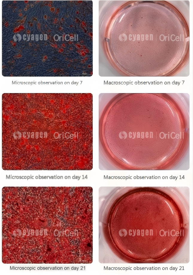
Step 4. Confirm Osteogenic Differentiation by Alizarin Red S Staining
Perform Alizarin Red S staining according to the following procedure to evaluate mineralization. The presence of red mineralized nodules indicates successful osteogenic differentiation.
- Remove the Induction Medium and Wash the Cells
Carefully aspirate the osteogenic differentiation medium and gently wash the cells 2–3 times with 1× PBS to remove any residual culture medium. - Fix the Cells
Add an appropriate volume of fixative to each well and incubate at room temperature for 30 minutes. - Perform Alizarin Red S Staining
Remove the fixative and wash the cells twice with 1× PBS. Add enough Alizarin Red S working solution to completely cover the cells and incubate at room temperature for 5–10 minutes. - Remove Excess Stain and Wash the Cells
Aspirate the staining solution and gently wash the cells 2–3 times with 1× PBS until no excess dye remains. Finally, add 1× PBS to keep the cells hydrated before microscopic observation.
Figure 1. Alizarin Red S staining results showing successful osteogenic differentiation and mineralized nodule formation. | Image credit: Cyagen OriCell
FAQ
Q: How do I know when to perform Alizarin Red S staining?
A: The timing of Alizarin Red S staining should not be determined solely by the duration of osteogenic induction. Instead, staining should be performed only after aggregated mineralized nodules are clearly visible under the microscope.
Q: What do mineralized nodules look like, and how can I identify them?
A: The images below show osteogenic differentiation of human bone marrow mesenchymal stem cells (left) and C57 mouse bone marrow mesenchymal stem cells (right).
The areas outlined in red indicate clearly visible mineralized nodules, which can be used as a reference when evaluating osteogenic differentiation prior to Alizarin Red S staining.
Q: What should I keep in mind when washing cells and selecting buffers for Alizarin Red S staining after osteogenic differentiation?
A: Avoid using calcium-containing buffers (such as PBS supplemented with Ca²⁺), as they may cause nonspecific binding and interfere with staining results.
After staining, wash the cells thoroughly to remove excess dye and minimize background staining.
Q: What should I do if the staining is too intense?
A: If the staining appears too intense, reduce the staining time or decrease the concentration of the Alizarin Red S solution (for example, to 0.1%).
Q: What should I do if the mineralized nodules detach during staining?
A: Handle the culture plate gently and avoid vigorous shaking before fixation to prevent the mineralized nodules from detaching.
Q: How should I select MSCs for osteogenic differentiation? Why are my cells failing to differentiate?
A: For optimal osteogenic differentiation, use low-passage MSCs that exhibit good morphology, high viability, and robust growth. Healthy cells with strong proliferative capacity are more likely to achieve successful osteogenic differentiation.
Q: What could cause poor Alizarin Red S staining results?
A: Several factors may contribute to unsatisfactory staining results:
Insufficient mineralized nodule formation
Before staining, confirm that osteogenic differentiation has been successfully induced. Staining is recommended only after abundant mineralized nodules are clearly visible under the microscope.
Staining solution temperature
Alizarin Red S solution is typically stored at 2–8°C. Allow the solution to equilibrate to room temperature before use.
Staining duration
The recommended staining time is 5–10 minutes. If the laboratory temperature is low or the staining solution is still cold, a longer incubation time may be required until satisfactory staining is observed.
Expired or improperly stored staining solution
If no staining develops even after prolonged incubation, the staining solution may have deteriorated due to improper storage or expiration and should be replaced.
OriCell Featured Products
| Type | Product Name | Cat. No. | Size |
|---|---|---|---|
| Cell Culture Media | OriCell Complete Medium For Human Bone Marrow Mesenchymal Stem Cells | HUXMA-90011 | 100 mL |
| Cell Culture Media | OriCell Human BMSC No-EXO Medium | HUXMA-90012 | 100 mL |
| Differentiation Medium | OriCell Osteogenic Differentiation Medium For Human Bone Marrow Mesenchymal Stem Cells | HUXMX-90021 | 100 mL |
Cyagen OriCell has long been dedicated to stem cell research, with professional expertise and rich experience in stem cell induced differentiation. Leveraging our well-established stem cell technology platform, we provide specialized reagents for osteogenic, adipogenic, and chondrogenic differentiation of various stem cells, featuring complete components and high differentiation efficiency. We also offer customized experimental services for stem cell differentiation potential studies.
About Cyagen OriCell
Cyagen OriCell is a Cyagen brand focused on the research and development of cell biology products, including stem cells, primary cells, and cell lines, as well as cell culture reagents and technical services. Serving universities, research institutes, hospitals, CROs, and CDMOs worldwide, Cyagen OriCell has accumulated extensive expertise in cell isolation and culture. The team has developed "spatial replication" culture technology to rapidly establish growth‑supportive environments, and runs an Antibiotic‑Free process grounded in strict environmental, materials, and personnel controls. Cyagen OriCell provides end‑to‑end solutions—from MSC isolation and identification to directed differentiation and assay services.
Cyagen OriCell's offerings are cited in over 10,000 publications, with a cumulative impact factor exceeding 90,000 and more than 160,000 citations, and the team has supported more than 3,000 research groups. Products are used by tens of thousands of customers across dozens of countries and regions.

