
Cell Culture Fungal Contamination: What It Is, Where It Comes From, and How to Fix It
When fungal contamination takes hold, mold hyphae can spread through the culture medium like unwanted cobwebs slowly overtaking a cleanroom, while yeast cells quietly turn the medium cloudy and fizzy—as if a tiny fermentation project had started in your incubator. Meanwhile, your cells, unaware of the takeover, are gradually becoming the main course.
In cell culture, contamination is a common laboratory problem that can severely compromise the reproducibility of experimental results and cell viability. So, how can fungal contamination be identified and prevented in routine practice? This article provides a detailed overview of the symptoms of fungal contamination and practical response strategies.
Q: What is fungal contamination in cell culture?
A: Fungal contamination in cell culture refers to the unintended introduction and proliferation of fungi, such as yeasts and molds, within the culture system during cell culture. This leads to deterioration of the cell culture environment and can severely compromise the purity, viability, metabolic function of cell lines, as well as the accuracy and reliability of experimental data.
Q: What factors may lead to fungal contamination in cell culture?
A: During cell culture, a lack of attention at any step may lead to fungal contamination.
1. Laboratory Environment (the most common and primary source of contamination)
- • Airflow and dust: Fungal spores, such as mold and yeast spores, are extremely small and lightweight, allowing them to remain suspended in the air for long periods. Personnel movement, opening and closing of doors, and ventilation systems in the laboratory can all generate airflow that carries spores from the external environment into the clean bench or cell culture room.
- • Humidity and cleanliness: If humidity in the cell culture room is too high or cleaning and disinfection are not thorough, damp areas such as wall corners and refrigerator door seals can easily support mold growth and become persistent sources of contamination.
- • Air-conditioning and ventilation systems: If central air-conditioning systems or ventilation ducts in the laboratory are not cleaned and maintained regularly, they can become reservoirs and disseminators of fungal spores.
2. Experimental Operations (the direct cause of contamination )
- • Attire: Entering the cell culture room without wearing proper cleanroom garments, or wearing outerwear that has been exposed to other contaminated areas, such as bacterial culture rooms or public corridors, may carry fungal spores.
- • Hand hygiene: Inadequate hand washing or insufficient disinfection, especially after touching shared items such as door handles, printers, and computers, can introduce contamination.
- • Speaking, coughing, and sneezing: These actions can release large amounts of aerosols inside the clean bench or cell culture room, and these aerosols may contain fungi.
- • Excessive or rapid movements: Moving arms or objects too quickly inside the clean bench can disrupt the unidirectional airflow and draw unfiltered external air into the work area.
3. Reagents and Consumables
- • Culture media and serum: Contamination during medium preparation, intrinsic contamination of serum, or improper storage of additives after opening may introduce fungi.
- • Dissociation enzymes and PBS: These can be easily contaminated if aliquoting or handling is improper.
- • Laboratory consumables: Culture flasks, reagent bottles, pipettes, and other consumables may also introduce contamination if their packaging is damaged or if sterilization during production is incomplete.
4. Contamination Originating from the Cells Themselves
- • Primary cells: Primary cells isolated from animal or human tissues may introduce fungi during handling.
- • Cross-contamination: Contaminated cells may transfer fungi to other cell lines during passaging or handling through shared reagents, pipettes, or culture medium.
5. Equipment and Facility Contamination
- • Water baths: Warm, stagnant water is a breeding ground for fungi and bacteria. If water baths are not cleaned and disinfected regularly, the risk of contamination is extremely high.
- • Refrigerators and water purification systems: Condensation water and water trays in 4°C refrigerators, as well as filter membranes and tubing in pure water systems that have not been maintained for long periods, may support microbial growth.
- • Microscopes and centrifuges: As shared equipment used by multiple people, microscopes and centrifuges can also become sources of contamination if cell samples leak and the surfaces are not cleaned and disinfected promptly.
Q: How can fungal contamination in cell culture be identified?
A: Fungal contamination in cell culture can be identified in several ways.
| Identification Method | Specific Characteristics |
|---|---|
| Visual inspection of the culture medium | Turbidity: The culture medium may appear cloudy, hazy, or flocculent. After standing, visible sediment or suspended aggregates may be observed. Colony morphology: White, pale yellow, black, or other flocculent, fluffy, or powdery colonies may be seen floating on the liquid surface or adhering to the bottom or side walls of the culture vessel. pH changes: In the early stage of contamination, pH changes in the culture medium may not be obvious, unlike bacterial contamination, which rapidly turns the medium yellow. However, as fungi proliferate extensively, the pH may also change. |
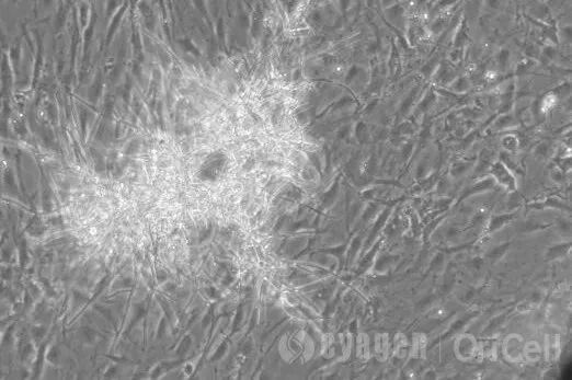
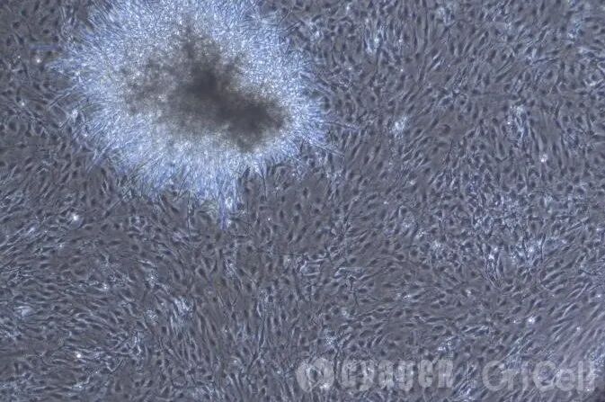
| Microscopic examination | Molds: Typical mycelia can be observed as slender, branched tubular structures that intertwine to form a network. Yeasts: Yeasts appear as individual oval or spherical cells, and budding reproduction can usually be observed. |
| Growth rate and pattern | Growth rate: Signs of contamination usually appear more slowly than those of bacterial contamination and are difficult to detect at the early stage. Growth pattern: Mold contamination often shows colony-like growth, gradually expanding outward from a single point; yeast contamination, by contrast, causes the medium to become uniformly turbid overall, with a cloudy appearance. |
| Effects on cells | Cell condition: Cell growth is significantly slowed, morphology deteriorates, intracellular granularity increases, and eventually, large numbers of cells die and detach. Nutrient competition: Fungi compete with cells for nutrients and growth factors in the culture medium. Metabolite toxicity: Metabolites secreted by fungi may alter the pH of the culture environment and exert toxic effects on cells. |
Typical fungal contamination cases
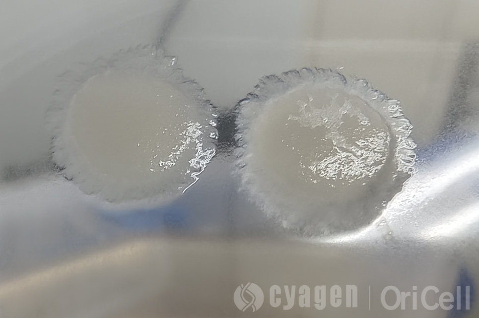
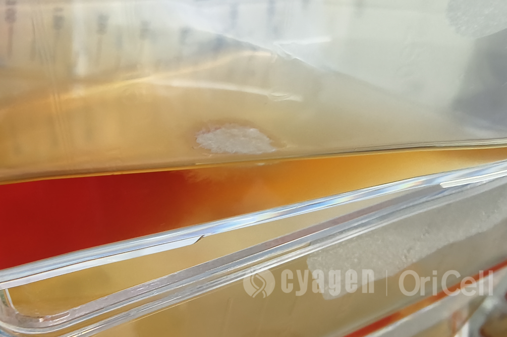
Typical fungal contamination cases under microscopy | Image credit: Cyagen OriCell
Q: How should cells affected by fungal contamination be handled?
A: In the vast majority of contamination cases, especially mold contamination, the cells should be discarded immediately and thorough disinfection should be performed. This is the safest, most economical, and most responsible approach for protecting other cells in the laboratory.
The operating steps are as follows:
- Immediately remove the contaminated culture from the incubator and clean bench, seal the flask cap with sealing film, and ensure that it is tightly sealed.
- For all contaminated cell flasks/dishes, culture medium, pipette tips that have come into contact with the culture, and related materials, if they are disposable consumables, discard all of them as biohazardous waste or perform high-temperature sterilization.
- Clean bench: Thoroughly wipe the work surface, inner walls, and all internal equipment with a chlorine-containing disinfectant or 75% ethanol. Then turn on the UV lamp for irradiation for more than 1 hour.
- Immediately empty the incubator, remove all other cells, and thoroughly wipe the inner walls, shelves, water tray, and all sensors of the incubator with a high-concentration disinfectant, such as a chlorine-containing disinfectant or a dedicated sporicidal agent.
- Check all reagents that may have been used during the operation, such as trypsin, PBS, and culture medium. For any opened reagent suspected of contamination, the safest approach is to sterilize it and then discard it.
- Replace the sterile water and clean the water tray regularly.
| Scenario | Recommended Solution |
|---|---|
| Most contamination cases, especially mold contamination | Immediate disposal and thorough disinfection. This is the safest, most economical, and most responsible approach for protecting other cells in the laboratory. |
| Unique or highly valuable contaminated cells | Rescue may be attempted under strict isolation using antifungal drug treatment, with multiple passages and repeated monitoring. However, it must be accepted that the success rate is low, the process is time-consuming and labor-intensive, and the treatment may have toxic effects on the cells themselves. After successful rescue, the cell line should also be cultured separately from other cells. |
OriCell Featured Products
| Type | Product Name | Cat. No. | Size |
|---|---|---|---|
| General Reagents | Penicillin-Streptomycin Solution (100X) | ATPS-10001 | 100 mL; 5 mL × 2 |
| General Reagents | Penicillin-Streptomycin-Amphotericin B Solution (100X) | APSB-10001 | 100 mL; 5 mL × 2 |
| General Reagents | Penicillin-Streptomycin-Gentamicin Solution (100X) | APSG-10001 | 100 mL; 5 mL × 2 |
| General Reagents | Mycoplasma Removal Agent (100X) | MPRA-10201 | 100 mL; 5 mL × 2 |
Cyagen OriCell provides a portfolio of cell culture reagents designed to support routine culture maintenance and contamination management. The products listed above include antibiotic solutions and mycoplasma removal reagents that can help researchers strengthen contamination control workflows while maintaining standardized cell culture practices.
About Cyagen OriCell
Cyagen OriCell is a Cyagen brand focused on the research and development of cell biology products, including stem cells, primary cells, and cell lines, as well as cell culture reagents and technical services. Serving universities, research institutes, hospitals, CROs, and CDMOs worldwide, Cyagen OriCell has accumulated extensive expertise in cell isolation and culture. The team has developed "spatial replication" culture technology to rapidly establish growth‑supportive environments, and runs an Antibiotic‑Free process grounded in strict environmental, materials, and personnel controls. Cyagen OriCell provides end‑to‑end solutions—from MSC isolation and identification to directed differentiation and assay services.
Cyagen OriCell's offerings are cited in over 10,000 publications, with a cumulative impact factor exceeding 90,000 and more than 160,000 citations, and the team has supported more than 3,000 research groups. Products are used by tens of thousands of customers across dozens of countries and regions.

