
 EN
EN
Establishing a Reliable HUVEC Angiogenesis Model: From Cell Preparation to Tube Formation
Establishing an in vitro angiogenesis model is essential for studying endothelial cell behavior, vascular development, and disease mechanisms. This is also a topic often discussed on Reddit and other lab forums, where researchers ask how to improve HUVEC tube formation, reduce variability between replicates, and select suitable culture and induction conditions. Among various endothelial cell models, human umbilical vein endothelial cells (HUVECs) are widely used due to their strong angiogenic capacity and relatively stable culture characteristics.
However, successful HUVEC angiogenesis experiments require careful optimization of multiple factors, including cell quality, induction conditions, culture environment, and experimental evaluation methods. Variations in these factors may affect endothelial tube formation and lead to inconsistent experimental outcomes.
To support researchers in establishing consistent angiogenesis models, OriCell provides a complete solution including high-quality HUVEC primary cells and optimized angiogenic induction medium.
The OriCell Human Umbilical Vein Endothelial Cell Angiogenesis Induction Medium Kit is designed to facilitate HUVEC angiogenic differentiation and support downstream evaluation of tube formation.
Let’s explore how OriCell solutions can support your vascular research journey.
Angiogenic Differentiation
HUVEC Angiogenic Induction and Tube Formation
Experimental Purpose
- • To study the in vitro angiogenic capacity of human umbilical vein endothelial cells.
- • To investigate the effects of different drugs on angiogenesis.
Experimental Principle
When endothelial cells are cultured on a specific matrix and stimulated by growth factors, integrin signaling pathways can be activated. Under these conditions, the cells mimic key steps of in vivo angiogenesis, including basement membrane degradation, followed by cell migration and eventual lumen formation.
Application Areas
- • Evaluate the effects of pro-angiogenic or anti-angiogenic drugs, genes, or signaling pathways.
- • Model tumor-associated angiogenesis in vitro.
- • Support the construction and optimization of vascularized tissue engineering models.
Experimental Materials and OriCell Product Solutions
| Category | OriCell Product / Material | Catalog Number | Application in This Assay |
|---|---|---|---|
| Primary cells | OriCell Human Umbilical Vein Endothelial Cells | HUVEC-20001 | Used as the primary endothelial cell model for angiogenic induction and tube formation assays. |
| Complete medium | OriCell Complete Medium For Human Umbilical Vein Endothelial Cells, regular type | HUVEC-90011 | Used for HUVEC recovery, maintenance culture, and expansion before induction. |
| Angiogenic induction reagent | OriCell Human Umbilical Vein Endothelial Cell Angiogenesis Induction Medium Kit, with Matrigel | HUVEC-90202 | Used for HUVEC angiogenic induction and tube formation experiments when Matrigel is included. |
| Angiogenic induction reagent | OriCell Human Umbilical Vein Endothelial Cell Angiogenesis Induction Medium Kit, without Matrigel | HUVEC-90201 | Used for HUVEC angiogenic induction and tube formation experiments when Matrigel is prepared separately. |
| Matrix reagent | OriCell Matrigel Matrix | MATR-10001 | Used to coat culture plates and provide the matrix environment required for endothelial tube formation. |
| Buffer | OriCell Phosphate-Buffered Saline Solution (1X) | PBS-10001 | Used for washing cells and related culture handling steps. |
| Dissociation reagent | OriCell Trypsin-EDTA Solution | TEDTA-10001 | Used for HUVEC dissociation during cell passaging and preparation before induction. |
Experimental Procedure
1. Cell Preparation
Cell recovery and culture: Recover cryopreserved HUVECs and culture them in HUVEC-specific complete medium until they reach 60%–70% confluence.
Cell pretreatment: Replace the medium with angiogenic induction medium and continue culture for 24 h.
Cell passaging: Digest the cells using 0.25% trypsin solution. When the cells become flattened and rounded, add culture medium to terminate digestion. Collect the cells, centrifuge, and resuspend them in angiogenic induction medium. Adjust the cell density to 1 × 10⁶ cells/mL.
2. Matrigel Plating
Matrigel thawing: Thaw Matrigel in advance in a 4°C refrigerator.
Plating: Evenly coat the pre-cooled culture wells with Matrigel. Equilibrate at 4°C for 5 min, then incubate at 37°C for 30 min to allow the Matrigel to solidify.
3. Cell Seeding
Cell seeding: For a 12-well plate, add 500 μL of induction medium to each well. Seed the prepared cell suspension onto the Matrigel at a density of 2–3 × 10⁵ cells/well.
Cell culture: Place the culture plate in a 37°C, 5% CO₂ incubator.
4. Observation and Imaging
Observation: Observe cell growth status and tubular structure formation under a microscope. In general, tube formation begins 2–4 h after seeding, reaches a peak at 4–10 h, and gradually shows tube disruption after 24 h.
Imaging: Capture images using a microscope to record tubular structure formation.
5. Quantitative Analysis
Tube structure quantification: Use image analysis software such as ImageJ to quantify total tube length, branch points, and the number of mesh-like network structures.
Statistical analysis: Use statistical software to analyze the data and compare differences among experimental groups.
HUVEC Angiogenic Induction Results
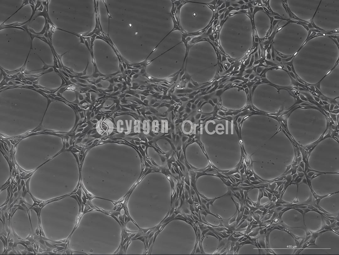
HUVEC-4 h, 50×
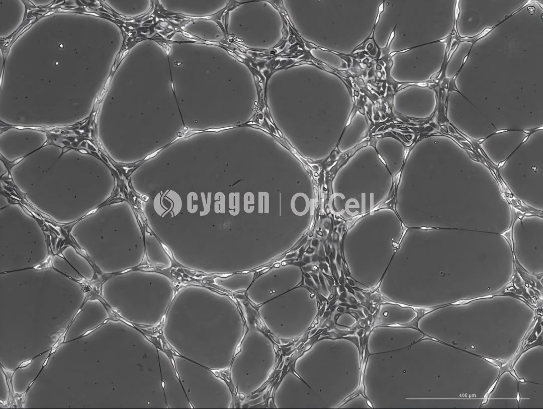
HUVEC-8 h, 50×
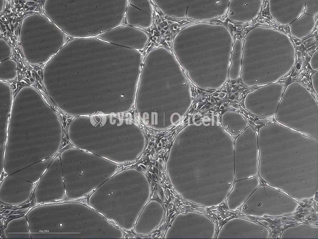
HUVEC-12 h, 50×
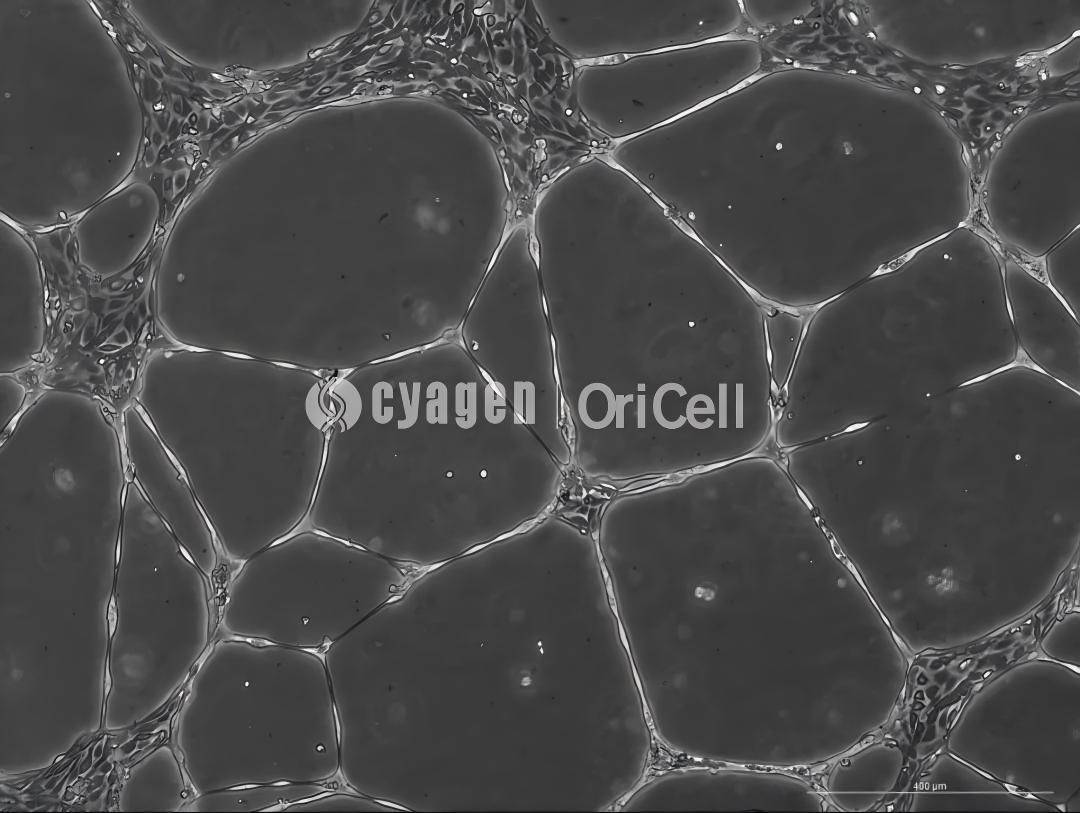
HUVEC-24 h, 50×
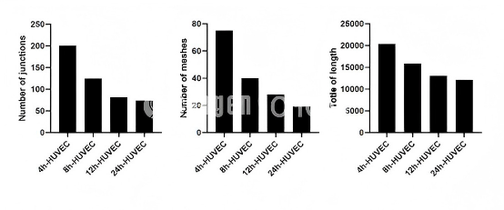
For tube formation assays, the final evaluation is usually based on three quantitative parameters: total length, junctions, and meshes. During HUVEC tube formation, cells are monitored and imaged at 4 h, 8 h, 12 h, and 24 h.
At 4 h of tube formation: Cells begin to form preliminary tubular structures. Connections appear between cells, but the tubular structures are still short and incomplete.
At 8 h of tube formation: Tubular structures extend further, branch points increase, and an initial network-like structure begins to form.
At 12 h of tube formation: Tubular structures become more complex, branch points and mesh-like structures increase significantly, and the lumens gradually become clearer.
At 24 h of tube formation: Tubular structures reach a peak and form a complete vessel-like network, although some tubular structures begin to break.
Summary: After human umbilical vein endothelial cells are seeded on Matrigel, tubular structures begin to form within 4 h, reach a peak at 12 h, and partially break after 24 h.
Experimental FAQ
Q1: Why must Matrigel be handled on ice?
A: Matrigel solidifies very easily at room temperature, so the entire operation should be performed on ice. Pipette tips, culture plates, and other consumables should also be pre-cooled before use.
Q2: Why should bubbles be avoided during Matrigel plating?
A: Bubbles must be avoided during plating, as they can seriously interfere with microscopic observation and affect image quality during tube formation evaluation.
Q3: What cell condition is required before HUVEC angiogenesis induction?
A: Cells should be in good condition before induction. In particular, excessive digestion during passaging should be avoided, as it may reduce cell viability and affect tube formation.
Q4: Why is aseptic technique important throughout the experiment?
A: Strict aseptic technique should be followed throughout the entire experiment to prevent cell contamination, which can compromise culture quality and experimental reliability.
Q5: How many technical replicates should be set for each group?
A: Each group should include three replicate wells to improve the accuracy, reproducibility, and reliability of the experimental data.
From HUVEC preparation to angiogenic differentiation and tube formation evaluation, establishing a reproducible workflow is essential for vascular research. With OriCell HUVEC products and optimized culture solutions, researchers can simplify experimental setup and improve consistency across studies.
About Cyagen OriCell
Cyagen OriCell is a Cyagen brand focused on the research and development of cell biology products, including stem cells, primary cells, and cell lines, as well as cell culture reagents and technical services. Serving universities, research institutes, hospitals, CROs, and CDMOs worldwide, Cyagen OriCell has accumulated extensive expertise in cell isolation and culture. The team has developed "spatial replication" culture technology to rapidly establish growth-supportive environments, and runs an Antibiotic-Free process grounded in strict environmental, materials, and personnel controls. Cyagen OriCell provides end-to-end solutions-from MSC isolation and identification to directed differentiation and assay services.
Cyagen OriCell's offerings are cited in over 10,000 publications, with a cumulative impact factor exceeding 90,000 and more than 160,000 citations, and the team has supported more than 3,000 research groups. Products are used by tens of thousands of customers across dozens of countries and regions.

