
 EN
EN
Can Any Tooth Be Used for DPMSC Isolation? Understanding Direct Explant and Enzymatic Digestion Methods
Dental pulp mesenchymal stem cells (DPMSCs), also known as dental pulp stem cells (DPSCs), are a valuable type of mesenchymal stem cell derived from dental pulp tissue. Since their discovery in healthy adult third molars, DPMSCs have attracted increasing attention in regenerative medicine research due to their strong proliferative capacity, multilineage differentiation potential, and relatively accessible tissue source. This is also a topic often discussed on Reddit and other lab forums, where researchers ask whether all tooth samples are suitable for DPMSC isolation and how to choose between direct explant culture and enzymatic digestion.
Compared with other mesenchymal stem cell sources, DPMSCs offer several advantages, including convenient tissue collection, low ethical concerns, and promising applications in areas such as dental tissue regeneration, periodontal research, and tissue engineering.
However, obtaining high-quality DPMSCs requires careful consideration of both tooth source selection and isolation strategy. Different isolation methods may influence cell outgrowth efficiency, initial cell recovery, and subsequent experimental performance.
Previously, we introduced a commonly used DPMSC isolation workflow based on dental pulp tissue processing. Read the related DPMSC isolation and culture guide. In this article, our scientists will further discuss two commonly used approaches for DPMSC isolation — the direct explant attachment method and the enzymatic digestion method — and explain how researchers can select an appropriate isolation strategy based on their experimental requirements.
So, can DPMSCs be isolated from any type of tooth? Let’s explore the factors that influence DPMSC isolation success.
Materials Preparation
Tissue samples: Healthy third molars without dental caries or naturally exfoliated deciduous teeth.
Experimental reagents: OriCell Phosphate-Buffered Saline Solution (1X), OriCell Complete Medium For Human Dental Pulp Mesenchymal Stem Cells, OriCell Trypsin Cell Dissociation Solution (0.25%), OriCell Collagenase Type I (0.1%), and OriCell Penicillin-Streptomycin Solution (100X).
Instruments and consumables: Cell culture dishes, ophthalmic forceps, dental forceps, and dental hammer.
Pretreatment of Extracted Teeth
Extracted teeth should be transported to the laboratory on ice, and dental pulp tissue should be collected within 12 h.
Rinse the extracted teeth twice with PBS containing 2% penicillin-streptomycin solution for 1 min each time. If dental caries are present, soak the tooth in 75% ethanol for 2–3 min before rinsing.
Dental Pulp Extraction
Place the rinsed extracted tooth in a culture dish containing PBS with 2% penicillin-streptomycin solution. The PBS should fully cover the tooth.
Use forceps to scrape away other tissues and impurities from the extracted tooth.
After all impurities are removed, transfer the tooth to a new culture dish. Use a dental hammer and dental forceps to separate the tooth body and expose the pulp cavity.
Transfer the tooth fragments to a new culture dish. Use ophthalmic forceps to peel out the dental pulp and place it in sterile PBS.
Isolation of Dental Pulp Mesenchymal Stem Cells
Direct Explant Attachment Method
Use ophthalmic forceps to tear the dental pulp into filament-like tissue fragments. Spread them evenly across the culture dish and allow them to air-dry naturally for 3–5 min.
During this period, prewarm OriCell Complete Medium For Human Dental Pulp Mesenchymal Stem Cells to 37°C.
After the dental pulp tissue adheres closely to the dish surface, gently add OriCell Complete Medium For Human Dental Pulp Mesenchymal Stem Cells along the wall of the dish.
Place the dish in a 37°C incubator with 5% CO₂ and 95% relative humidity for culture.
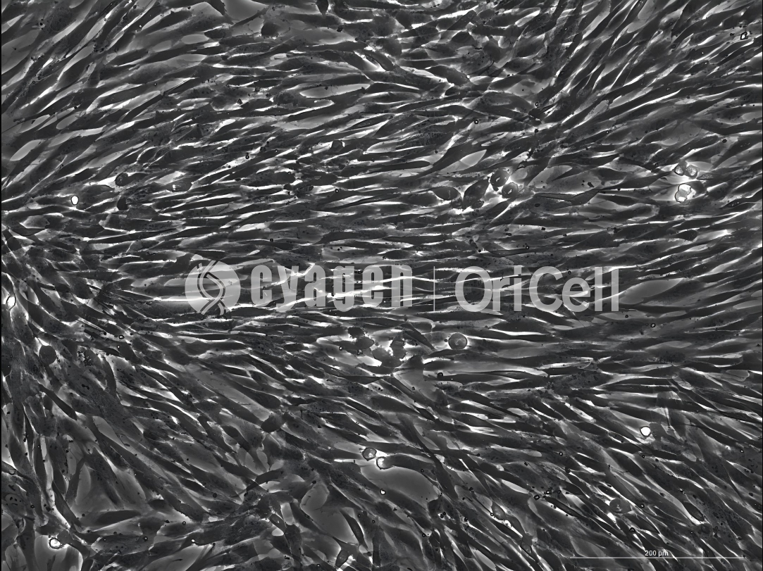
During the first 72 h of culture, avoid moving the culture dish as much as possible. After 72 h, observe cell migration under a microscope and change the medium as needed. When cell confluence reaches 80%–90%, the cells can be passaged.
Enzymatic Digestion Method
Prewarm 0.1% Collagenase Type I to 37°C.
Cut the dental pulp tissue into fragments of approximately 1 mm². Collect the tissue fragments in a centrifuge tube and centrifuge at 265 × g for 5 min at room temperature.
Discard the supernatant. Resuspend the dental pulp tissue in 5 mL of 0.1% Collagenase Type I and digest in a 37°C water bath for 20 min. During digestion, take out the tube every 5 min and mix thoroughly. A thermostatic shaker can also be used if available.
After digestion, add an equal volume of complete medium and mix the digestion suspension thoroughly with the complete medium.
Centrifuge at 265 × g for 5 min and discard the supernatant. Resuspend the cells in prewarmed OriCell Complete Medium For Human Dental Pulp Mesenchymal Stem Cells, then seed the cells into a culture dish.
After seeding, avoid moving the culture dish as much as possible. Observe cell status after 72 h, and change the medium as needed. When cell confluence reaches 80%–90%, the cells can be passaged.
Passaging of Dental Pulp Mesenchymal Stem Cells
Human dental pulp mesenchymal stem cells should be passaged when they grow to 80%–90% confluence. Do not allow the cells to reach complete or excessive confluence, as this may cause contact inhibition and severely affect cell growth.
Passaging Procedure
- Prewarm the complete medium and 0.25% trypsin to 37°C.
- Aspirate the culture medium. Wash the cells 2–3 times with 1× PBS to remove residual serum, then aspirate the PBS.
- Add an appropriate amount of 0.25% trypsin and gently rotate the culture dish to allow the trypsin to cover the cell surface. Digest for 1–2 min. Under the microscope, the cell gaps should become larger and the cells should become rounded. The digestion endpoint should be determined based on microscopic observation.
- Gently tap the wall of the culture vessel by hand. When approximately 80% of the cells detach and float, immediately add 2–3 mL of complete medium to terminate digestion.
- Transfer the cells into a centrifuge tube. Add 1× PBS to the culture flask and wash 1–2 times, then transfer the wash solution to the same centrifuge tube.
- Centrifuge at 265 × g for 4 min. Aspirate the supernatant and resuspend the cells in 2–3 mL of complete medium.
- Seed the cells according to the cell number or at a density of 2 × 10⁴ cells/cm².
- Add complete medium and culture the cells in a 37°C incubator with 5% CO₂ and 95% relative humidity.
FAQ
Q1: Can both the direct explant attachment method and the enzymatic digestion method be used to isolate DPMSCs?
A: Yes. Both the direct explant attachment method and the enzymatic digestion method can be used to obtain dental pulp mesenchymal stem cells. However, the choice of method should be based on the tooth source and tissue condition.
Q2: Which method is recommended for deciduous teeth?
A: If the experimental material is a deciduous tooth, the direct explant attachment method is recommended to increase cell yield.
Q3: Why is the direct explant attachment method preferred for deciduous teeth?
A: Dental pulp from deciduous teeth is usually fragile, soft, and small in volume. The direct explant attachment method helps minimize operation time and avoids excessive handling steps, reducing additional loss of dental pulp tissue.
Q4: How soon should dental pulp tissue be extracted after tooth collection?
A: Extracted teeth should be transported on ice and processed within 12 h for dental pulp extraction.
Q5: Why should the culture dish not be moved during the first 72 h after seeding?
A: In both direct explant culture and enzymatic digestion culture, minimizing dish movement during the first 72 h helps tissue fragments or newly seeded cells remain stable, which supports cell attachment and outgrowth.
Q6: When should human dental pulp mesenchymal stem cells be passaged?
A: Human dental pulp mesenchymal stem cells should be passaged when cell confluence reaches 80%–90%.
Q7: Why should DPMSCs not be allowed to become completely confluent or overconfluent?
A: Complete or excessive confluence may lead to contact inhibition, which can severely affect cell growth.
Q8: How should the digestion endpoint be determined during passaging?
A: The digestion endpoint should be determined under a microscope. When cell gaps become larger, cells become rounded, and approximately 80% of the cells detach after gentle tapping, complete medium should be added immediately to terminate digestion.
Q9: Why should the total digestion time not be too long?
A: For mesenchymal stem cells, the time required for enzymatic detachment is usually short. Cells that are difficult to detach by digestion may mostly be non-mesenchymal stem cells or other contaminating cell types.
Complete OriCell Solution for Dental Pulp Mesenchymal Stem Cells
With 17 years of experience in stem cell culture, Cyagen OriCell provides a complete DPMSC solution to support dental pulp mesenchymal stem cell research.
In addition, Cyagen OriCell also provides complete solutions for mesenchymal stem cells derived from bone marrow, adipose tissue, umbilical cord, and umbilical cord blood. Please contact us for consultation or ordering information.
References
- Gronthos S, Mankani M, Brahim J, et al. Postnatal human dental pulp stem cells (DPSCs) in vitro and in vivo. Proceedings of the National Academy of Sciences of the United States of America. 2000;97(25):13625–13630.
- Zhongyuan Union Cell & Gene Engineering Co., Ltd. Announcement on a drug candidate of an investee subsidiary entering Phase II clinical trial. Announcement No. 2023-022, May 25, 2023.

