
 EN
EN
Black Dots in Cell Culture: Causes, Identification, and Troubleshooting Guide
The appearance of unexpected black dots or particles in cell culture is a common concern among researchers. These mysterious structures observed under the microscope may originate from various sources, including cell debris, cellular secretions, precipitates from culture components, microscope-related artifacts, or potential microbial contamination.
Although some black particles may be harmless, others can indicate underlying issues that affect cell growth, morphology, viability, and experimental reproducibility. Therefore, accurately identifying the cause of these particles is essential for maintaining reliable cell culture conditions and ensuring high-quality research results.
In this article, we will explore the common causes of black dots in cell culture, how to distinguish between different types of particles through microscopic observation and experimental evaluation, and the appropriate troubleshooting strategies. By understanding these potential issues, researchers can better maintain their cell cultures and minimize unexpected disruptions during experiments.
Potential Cause 1: Cell Debris Associated with Cellular Aging and Apoptosis
As passage number increases, particularly in proliferative primary cells such as mesenchymal stem cells, cells may show reduced proliferation, altered morphology, and an increased number of small black particles between cells.
Recommended approach: Use low-passage cells whenever possible. For mesenchymal stem cells, low-passage cells are recommended for experiments such as trilineage differentiation, drug treatment, and molecular characterization.
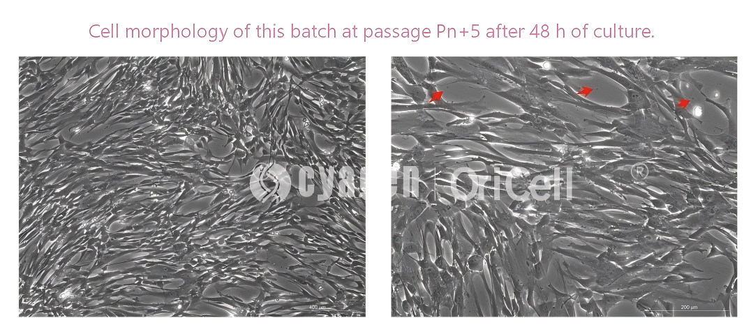
Using OriCell human umbilical cord mesenchymal stem cells as an example, a small number of black particles may be observed under the microscope as passage number increases. This is generally considered a normal phenomenon and does not affect use, although low-passage cells are still recommended for research applications whenever possible.
Potential Cause 2: Suboptimal Handling During Cell Passaging
Customer case: OriCell human umbilical cord mesenchymal stem cells cultured by a customer are used here as an example.
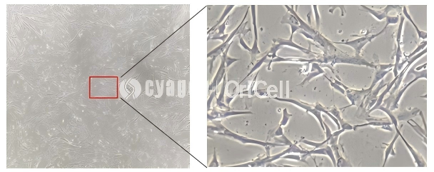
Left image: Under low magnification, the cell morphology appears to have changed.
Right image: Upon closer observation, the cells show a stretched or filamentous appearance, with numerous small black particles present in the spaces between cells.
Possible causes identified: Delayed passaging and excessive digestion time.
Practical tip: When purchasing cells, researchers may consult the cell supplier regarding the recommended passaging time for the specific cell type. Passaging should be adjusted proportionally based on the actual growth status of each batch and passage.
Potential Cause 3: Contamination or Staining on the Eyepiece or Objective Lens
This cause is relatively easy to distinguish. If the culture flask or culture dish is moved under the microscope and the black dots do not move accordingly, the particles are likely caused by dirt or staining on the eyepiece or objective lens.
The eyepiece or objective lens can be gently removed and carefully wiped with lens paper moistened with a small amount of anhydrous ethanol or a dedicated lens-cleaning solution to remove surface contamination.
Note: These cleaning solutions are flammable and should be handled with caution.
Potential Cause 4: Microbial Contamination
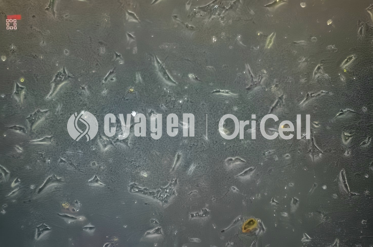
When cells are contaminated with bacteria, fungi, or mycoplasma, the number of small black particles may increase rapidly. This phenomenon is especially obvious in cases of bacterial contamination.
As shown in the customer-provided results below, on day 1, a small number of floating cells can be seen among the cell population, while the black particles are barely visible without careful observation. By day 2, however, under low magnification, the number of black particles between cells has increased markedly, cell morphology has already changed, and the phenomenon is even more evident under higher magnification. In addition, these black particles may show active motility.
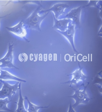
Recommended approach: Discard the contaminated culture directly. For any microbial contamination, the earlier the culture is discarded, the lower the overall loss. If the cell line is exceptionally valuable, penicillin-streptomycin solution or even a broader antibiotic combination such as penicillin-streptomycin-gentamicin may be added to the medium as a temporary measure to maintain the cells, but samples should be collected as soon as possible. During subsequent culture, strict precautions are also required to prevent contamination of other cells or culture media. The presence of antibiotics in the culture medium does not mean that cells are protected from contamination.
OriCell Featured Products
| Type | Product Name | Cat. No. | Size |
|---|---|---|---|
| General Reagents | Penicillin-Streptomycin Solution (100X) | ATPS-10001 | 100 mL; 5 mL × 2 |
| General Reagents | Penicillin-Streptomycin-Amphotericin B Solution (100X) | APSB-10001 | 100 mL; 5 mL × 2 |
| General Reagents | Penicillin-Streptomycin-Gentamicin Solution (100X) | APSG-10001 | 100 mL; 5 mL × 2 |
| General Reagents | Mycoplasma Removal Agent (100X) | MPRA-10201 | 100 mL; 5 mL × 2 |
Cyagen OriCell provides a portfolio of cell culture reagents designed to support routine culture maintenance and contamination management. The products listed above include antibiotic solutions and mycoplasma removal reagents that can help researchers strengthen contamination control workflows while maintaining standardized cell culture practices.
About Cyagen OriCell
Cyagen OriCell is a Cyagen brand focused on the research and development of cell biology products, including stem cells, primary cells, and cell lines, as well as cell culture reagents and technical services. Serving universities, research institutes, hospitals, CROs, and CDMOs worldwide, Cyagen OriCell has accumulated extensive expertise in cell isolation and culture. The team has developed "spatial replication" culture technology to rapidly establish growth‑supportive environments, and runs an Antibiotic‑Free process grounded in strict environmental, materials, and personnel controls. Cyagen OriCell provides end‑to‑end solutions—from MSC isolation and identification to directed differentiation and assay services.
Cyagen OriCell's offerings are cited in over 10,000 publications, with a cumulative impact factor exceeding 90,000 and more than 160,000 citations, and the team has supported more than 3,000 research groups. Products are used by tens of thousands of customers across dozens of countries and regions.

