
Chondrogenic Differentiation Medium For Rat Adipose-derived Mesenchymal Stem Cells
Introduction
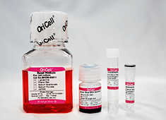
The OriCell™ Chondrogenic Differentiation Medium For Rat Adipose-derived Mesenchymal Stem Cells, developed by our R&D team, contains a basic medium and supplements suitable for the growth of rat adipose-derived mesenchymal stem cells. Extensive cell culture data have demonstrated that this product can induce these cells to differentiate into cartilage tissue stably and efficiently.
When citing our products in academic journals, please indicate "OriCell™ + Catalog Number, from Cyagen Biosciences (Guangzhou) Inc."
Product Information
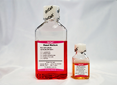
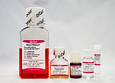
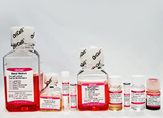
| Components | Catalog Number | Volume |
| OriCell™ Basal Medium For Cell Culture | BHDM-03011 | 97 mL |
| OriCell™ Supplement For Rat Adipose-derived Mesenchymal Stem Cells Chondrogenic Differentiation I | RAXMD-04041-1 | 2 mL |
| OriCell™ Supplement For Rat Adipose-derived Mesenchymal Stem Cells Chondrogenic Differentiation II | RAXMD-04041-2 | 1 mL |
|
OriCell™ Alcian Blue 8GX Solultion (pH=2.1~2.3) |
ALCB-10001 | 5 mL |
QC
- Pass the detection of bacteria, fungi, mycoplasma and endotoxins.
- Pass the detection of osmotic pressure and pH.
- Pass the detection of product quality.
Please refer to "COA" for details.
General Handing Principles
- Ensure that all equipment is kept clean and tidy.
- Standard operation method. Please operate according to the method described in the product manual.
- The ingredients should be properly stored in accordance with the storage conditions and used as soon as possible.
- If the complete medium cannot be used up within a short period, prepare it in batches according to the volume ratio of each component in the kit, and store in aliquots for future use.
Product Stability and Storage Conditions
- All ingredients must be kept in dark place.
- The basal medium should be stored in a refrigerator at 4°C for a period of 1 year. Other components should be stored at -20°C for a period of 2 years.
- The prepared complete medium can be stored at 4°C for up to 1 month. If culture conditions remain stable, the container is well-sealed, and temperature fluctuations are avoided, the storage period may be extended appropriately but should not exceed 45 days.
- Please use all products before the expiration date. Expired ingredients may significantly affect the cell culture effect.
Note: This product is intended for research use only and is not for diagnostic, therapeutic,
clinical, household, or any other applications.
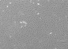
Wistar Rat Adipose-derived Mesenchymal Stem Cells
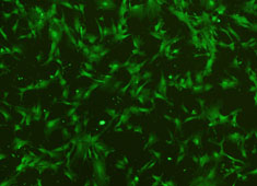
SD Rat Adipose-derived Mesenchymal Stem Cells With GFP
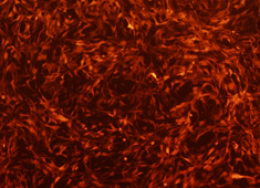
SD Rat Adipose-derived Mesenchymal Stem Cells With RFP

Complete Medium For Rat Adipose-derived Mesenchymal Stem Cells
Complete Medium For Rat Adipose-derived Mesenchymal Stem Cells (Without EXO)
Osteogenic Differentiation Medium For Rat Adipose-derived Mesenchymal Stem Cells
Adipogenic Differentiation Medium For Rat Adipose-derived Mesenchymal Stem Cells
SD Rat Adipose-derived Mesenchymal Stem Cell Exosomes
If you are interested in our products or services, you can contact us using
the methods below.
Submit your requests
Email us via [email protected]

