Chondrogenic Differentiation Medium For Bone Marrow Mesenchymal Stem Cells (Multipurpose)
Introduction
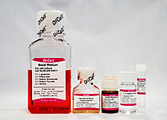
OriCell™ Chondrogenic Differentiation Medium For Bone Marrow Mesenchymal Stem Cells (Multipurpose) is specially formulated and developed by our R&D team. It consists of an optimized basal medium and essential supplements, tailored to induce the chondrogenic differentiation of bone marrow mesenchymal stem cells (BMSCs).
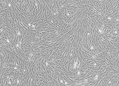
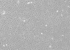
This product is suitable for chondrogenic induction and differentiation of BMSCs from rabbits, canines, monkeys, and other species. Extensive data have demonstrated that it can efficiently and stably induce these cells to differentiate into cartilage tissue.
When citing our products in academic publications, please use the following format: "OriCell™ [Product Name] + [Catalog Number], from Cyagen Biosciences."
Product Information
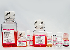
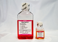
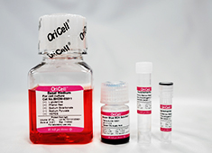
| Components | Catalog Number | Volume | Storage |
| OriCell™ Basal Medium For Cell Culture | BHDM-03011 | 97 mL | 4 °C |
| OriCell™ Supplement For Bone Marrow Mesenchymal Stem Cells Chondrogenic Differentiation I | GUXMX-04041-1 | 2 mL | -20 °C |
| OriCell™ Supplement For Bone Marrow Mesenchymal Stem Cells Chondrogenic Differentiation II | GUXMX-04041-2 | 1 mL | -20 °C |
| OriCell™ Alcian Blue 8GX Solultion (pH=2.1~2.3) | ALCB-10001 | 5 mL | 4 °C |
QC
- Pass the detection of bacteria, fungi, mycoplasma and endotoxins.
- Pass the detection of osmotic pressure and pH.
- Pass the detection of product quality.
Please refer to "COA" for details.
General Handling Principles
- Maintain strict aseptic technique. Ensure complete sterility throughout all procedures, particularly within the laminar flow hood and incubator.
- Follow standardized protocols. Adhere strictly to the product manual instructions. Implement rigorous control over experimental variables and include appropriate parallel controls.
- Ensure proper storage and use. Store all components according to specified conditions and use them promptly to ensure optimal performance.
- Aliquot for long-term storage. If the entire volume will not be used up immediately, prepare the medium in batches according to the specified component volume ratios and store in aliquots.
Product Stability and Storage Conditions
- All components must be stored away from light.
- The basal medium and Alcian Blue 8GX solultion have a shelf life of 1 year and should be stored at 4 °C. Other components have a shelf life of 2 years and should be stored at -20 °C.
- Once prepared, the medium has a shelf life of 1 month when stored at 4 °C. The shelf life may be extended up to a maximum of 45 days, provided that culture conditions remain stable, the container is properly sealed, and repeated temperature fluctuations are avoided.
- Use all components before the expiration dates. Expired components may severely compromise culture performance.
- Canine bone marrow mesenchymal stromal cells with lentiviral mHCN4 gene transfer create cardiac pacemakers. 点击索取文献 ››
- Change in Hepatocyte Growth Factor Concentration Promote Mesenchymal Stem Cell-Mediated Osteogenic Regeneration. 点击索取文献 ››
- Intervertebral disc regeneration using platelet‑rich plasma‑containing bone marrow‑derived mesenchymal stem cells A preliminary investigation. 点击索取文献 ››
- Directing the osteoblastic and chondrocytic differentiations of mesenchymal stem cells: matrix vs. induction media. 点击索取文献 ››
- The potential of using semitendinosus tendon as autograft in rabbit meniscus reconstruction. 点击索取文献 ››
- Pericellular collagen I coating for enhanced homing and chondrogenic differentiation of mesenchymal stem cells in direct intra-articular injection. 点击索取文献 ››
- Is exclusion of leukocytes from platelet-rich plasma (PRP) a better choice for early intervertebral disc regeneration?. 点击索取文献 ››
- Co-delivery of Synovium-derived Mesenchymal Stem Cells and TGF-# by a Hybrid Scaffold for Cartilage Regeneration. 点击索取文献 ››
- Hypoxic mesenchymal stem cell-derived exosomes promote bone fracture healing by the transfer of miR-126. 点击索取文献 ››
-
Canine bone marrow mesenchymal stromal cells with lentiviral mHCN4 gene transfer create cardiac pacemakers.
Cytotherapy 14:529 (2012) IF = 3.293 Year 2012
-
Change in Hepatocyte Growth Factor Concentration Promote Mesenchymal Stem Cell-Mediated Osteogenic Regeneration.
J Cell Mol Med 16:1260 (2012) IF = 4.014 Year 2012
-
Intervertebral disc regeneration using platelet‑rich plasma‑containing bone marrow‑derived mesenchymal stem cells A preliminary investigation.
Mol Med Rep 13:3475 (2016) IF = 1.922 Year 2016
-
Directing the osteoblastic and chondrocytic differentiations of mesenchymal stem cells: matrix vs. induction media.
Regenerative Biomaterials 4:269-279 (2017) Year 2017
-
The potential of using semitendinosus tendon as autograft in rabbit meniscus reconstruction.
Scientific RepoRts 7:7033 (2017) IF = 4.259 Year 2017
-
Pericellular collagen I coating for enhanced homing and chondrogenic differentiation of mesenchymal stem cells in direct intra-articular injection.
Stem Cell Res Ther 9:174 (2018) IF = 4.963 Year 2018
-
Is exclusion of leukocytes from platelet-rich plasma (PRP) a better choice for early intervertebral disc regeneration?.
Stem Cell Res Ther 9 (1):199 (2018) IF = 4.963 Year 2018
-
Co-delivery of Synovium-derived Mesenchymal Stem Cells and TGF-# by a Hybrid Scaffold for Cartilage Regeneration.
ACS Biomater. Sci. Eng. DOI:10.1021/acsbiomaterials.8b00483 (2018) IF = 4.432 Year 2018
-
Hypoxic mesenchymal stem cell-derived exosomes promote bone fracture healing by the transfer of miR-126.
Acta Biomaterialia PMID: 31857259 (2019) IF = 6.638 Year 2019