
Chondrogenic Differentiation Medium For Mouse Bone Marrow Mesenchymal Stem Cells
Introduction
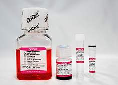
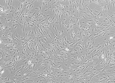
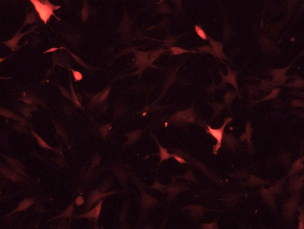
The OriCell™ Chondrogenic Differentiation Medium For Mouse Bone Marrow Mesenchymal Stem Cells, carefully developed by the OriCell™ R&D team, contains a basic medium and various additives which are suitable for the growth of mouse bone marrow mesenchymal stem cells. Extensive cell culture data have demonstrated that this product can stably and efficiently induce these cells to differentiate into cartilage tissue.
When citing our products in academic journals, please indicate "OriCell™ + Catalog Number, from Cyagen Biosciences (Guangzhou) Inc."
Product Information
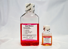
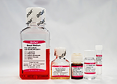
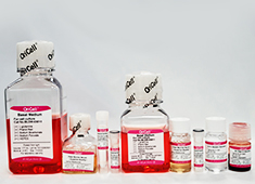
| Components | Catalog Number | Volume |
| OriCell™ Basal Medium For Cell Culture | BHDM-03011 | 97 mL |
| OriCell™ Supplement For Mouse Bone Marrow Mesenchymal Stem Cells Chondrogenic Differentiation I | MUXMX-04041-1 | 2 mL |
| OriCell™ Supplement For Mouse Bone Marrow Mesenchymal Stem Cells Chondrogenic Differentiation II | MUXMX-04041-2 | 1 mL |
| OriCell™ Alcian Blue 8GX Solution (pH=2.1~2.3) | ALCB-10001 | 5 mL |
QC
- Pass the detection of bacteria, fungi, mycoplasma and endotoxins.
- Pass the detection of osmotic pressure and pH.
- Pass the detection of product quality.
Please refer to "COA" for details.
General Handing Principles
- Ensure that all equipment is kept clean and tidy.
- Standard operation method. Please operate according to the method described in the product manual.
- The ingredients should be properly stored in accordance with the storage conditions and used as soon as possible.
- If the complete medium cannot be used up within a short period, prepare it in batches according to the volume ratio of each component in the kit, and store in aliquots for future use.
Product Stability and Storage Conditions
- All ingredients must be kept in dark place.
- The basal medium should be stored in a refrigerator at 4°C for a period of 1 year. Other components should be stored at -20°C for a period of 2 years.
- The prepared complete medium can be stored at 4°C for up to 1 month. If culture conditions remain stable, the container is well-sealed, and temperature fluctuations are avoided, the storage period may be extended appropriately but should not exceed 45 days.
- Please use all products before the expiration date. Expired ingredients may significantly affect the cell culture effect.
Note: This product is intended for research use only and is not for diagnostic, therapeutic,
clinical, household, or any other applications.
- Implantation of biomaterial as an efficient method to harvest mesenchymal stem cells. 点击索取文献 ››
- MicroRNA-145 Regulates Chondrogenic Differentiation of Mesenchymal Stem Cells by Targeting Sox9. 点击索取文献 ››
- Activation of canonical wnt pathway promotes differentiation of mouse bone marrow-derived MSCs into type II alveolar epithelial cells, confers resistance to oxidative stress, and promotes their migration to injured lung tissue in vitro. 点击索取文献 ››
- Migration of CXCR4 Gene‐Modified Bone Marrow‐Derived Mesenchymal Stem Cells to the Acute Injured Kidney. 点击索取文献 ››
- Allogeneic compact bone-derived mesenchymal stem cell transplantation increases survival of mice exposed to lethal total body irradiation: a potential immunological mechanism. 点击索取文献 ››
- Bone marrow mesenchymal stem cells promote the repair of islets from diabetic mice through paracrine actions. 点击索取文献 ››
- Mesenchymal Stem Cells Promote Diabetic Corneal Epithelial Wound Healing Through TSG-6–Dependent Stem Cell Activation and Macrophage Switch. 点击索取文献 ››
- Intramembranous ossification and endochondral ossification are impaired differently between glucocorticoid-induced osteoporosis and estrogen deficiency-induced osteoporosis. 点击索取文献 ››
- Simultaneous isolation of mesenchymal stem cells and endothelial progenitor cells derived from murine bone marrow. 点击索取文献 ››
- Enhanced alleviation of aGVHD by TGF-β1-modified mesenchymal stem cells in mice through shifting MΦ into M2 phenotype and promoting the differentiation of Treg cells. 点击索取文献 ››
- Exosome-shuttled miR-216a-5p from hypoxic preconditioned mesenchymal stem cells repair traumatic spinal cord injury by shifting microglial M1/M2 polarization. 点击索取文献 ››
- Extracellular vesicles from bone marrow mesenchymal stem cells alleviate acute rejection injury after liver transplantation by carrying miR-22-3p and inducing M2 polarization of Kupffer cells. 点击索取文献 ››
-
Implantation of biomaterial as an efficient method to harvest mesenchymal stem cells.
Exp Biol Med (Maywood) 236:1477 (2011) IF = 2.165 Year 2011
-
MicroRNA-145 Regulates Chondrogenic Differentiation of Mesenchymal Stem Cells by Targeting Sox9.
PLoS One 21799743 (2011) IF = 3.234 Year 2011
-
Activation of canonical wnt pathway promotes differentiation of mouse bone marrow-derived MSCs into type II alveolar epithelial cells, confers resistance to oxidative stress, and promotes their migration to injured lung tissue in vitro.
J Cell Physiol 228:1270 (2013) IF = 3.874 Year 2013
-
Migration of CXCR4 Gene‐Modified Bone Marrow‐Derived Mesenchymal Stem Cells to the Acute Injured Kidney.
J Cell Biochem 114:2677 (2013) IF = 3.263 Year 2013
-
Allogeneic compact bone-derived mesenchymal stem cell transplantation increases survival of mice exposed to lethal total body irradiation: a potential immunological mechanism.
Chin Med J (Engl) 127:475 (2014) Year 2014
-
Bone marrow mesenchymal stem cells promote the repair of islets from diabetic mice through paracrine actions.
Mol Cell Endocrinol 388:41 (2014) IF = 4.405 Year 2014
-
Mesenchymal Stem Cells Promote Diabetic Corneal Epithelial Wound Healing Through TSG-6–Dependent Stem Cell Activation and Macrophage Switch.
Investigative Ophthalmology & Visual Science 5810:4064-4074 (2017) IF = 3.303 Year 2017
-
Intramembranous ossification and endochondral ossification are impaired differently between glucocorticoid-induced osteoporosis and estrogen deficiency-induced osteoporosis.
Sci Rep 8:3867 (2018) IF = 4.259 Year 2018
-
Simultaneous isolation of mesenchymal stem cells and endothelial progenitor cells derived from murine bone marrow.
Exp Ther Med 16:5171-5177 (2019) IF = 1.41 Year 2019
-
Enhanced alleviation of aGVHD by TGF-β1-modified mesenchymal stem cells in mice through shifting MΦ into M2 phenotype and promoting the differentiation of Treg cells.
JOURNAL OF CELLULAR AND MOLECULAR MEDICINE PMID: 31782262 (2019) IF = 4.658 Year 2019
-
Exosome-shuttled miR-216a-5p from hypoxic preconditioned mesenchymal stem cells repair traumatic spinal cord injury by shifting microglial M1/M2 polarization.
Journal of Neuroinflammation DOI: 10.1186/s12974-020-1726-7 (2020) IF = 5.7 Year 2020
-
Extracellular vesicles from bone marrow mesenchymal stem cells alleviate acute rejection injury after liver transplantation by carrying miR-22-3p and inducing M2 polarization of Kupffer cells.
JOURNAL OF GENE MEDICINE IF = 4.152 Year 2023

C57BL/6 Mouse Bone Marrow Mesenchymal Stem Cells
Balb/c Mouse Bone Marrow Mesenchymal Stem Cells

C57BL/6 Mouse Bone Marrow Mesenchymal Stem Cells With GFP
Balb/c Mouse Bone Marrow Mesenchymal Stem Cells With GFP
C57BL/6 Mouse Bone Marrow Mesenchymal Stem Cells With RFP
Balb/c Mouse Bone Marrow Mesenchymal Stem Cells With RFP

Complete Medium For Mouse Bone Marrow Mesenchymal Stem Cells
Complete Medium For Mouse Bone Marrow Mesenchymal Stem Cells (Without EXO)
Osteogenic Differentiation Medium For Mouse Bone Marrow Mesenchymal Stem Cells
Adipogenic Differentiation Medium For Mouse Bone Marrow Mesenchymal Stem Cells
If you are interested in our products or services, you can contact us using
the methods below.
Submit your requests
Email us via [email protected]

